This article has been reviewed according to Science X's editorial process and policies. Editors have highlighted the following attributes while ensuring the content's credibility:
fact-checked
proofread
Demonstrating fetal head moldability and brain compression

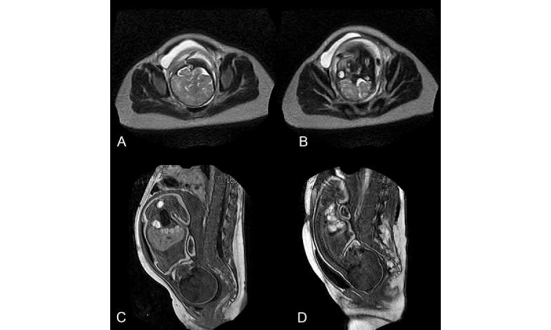
Researchers have called for improvements in birthing risk assessment and anticipatory interventions. In a study titled "Using Magnetic Resonance Imaging During Childbirth to Demonstrate Fetal Head Moldability and Brain Compression: Prospective Cohort Study," published in JMIR Formative Research, they observe that the birthing process is difficult to assess using simple imaging technology because the maternal bony pelvis and fetal skeleton interfere with visualizing the soft tissues.
Polygonal meshes for each part of the fetal body were used to study fetal head moldability and brain compression. In this case, observable brain shape deformation demonstrated that brain compression had occurred and was not necessarily well tolerated by the fetus.
Depending on fetal head moldability, these observations suggest that cephalopelvic disproportion can result in either obstructed labor or major fetal head molding with brain compression. MRI might be the best imaging technology to explore all combined aspects of cephalopelvic disproportion and achieve a better understanding of the underlying mechanisms of fetal head molding and moldability.
Dr. Olivier Ami from the Clinique de la Muette—Ramsay Santé said, "Events occurring within the first few hours of birth can cause fetal cerebral palsy or maternal perineum trauma, determining a human's life trajectory."
Magnetic resonance imaging is a noninvasive and nonirradiating tool suitable for exploring the biomechanics of the birthing process and evaluating fetal well-being. A clinical protocol to achieve this goal requires numerous safeguards to ensure the same level of safety in the MRI suite as in the delivery room.
In 1948, Mengert described five components of cephalopelvic disproportion:
- Size and shape of the bony pelvis,
- Size of the fetal head,
- Force exerted by the uterine contractions,
- Moldability of the fetal head, and
- Presentation and position of the fetal head.
Exploring these factors with imaging during birth is crucial to the prediction and prevention of cephalopelvic disproportion.
MRI is routinely used to explore fetal malformations antenatally and perform obstetric pelvimetry. However, despite the potential advantages of MRI when evaluating the birthing process in real time, these authors identified only a few sites worldwide with an open-field MRI near a maternity ward that would enable the exploration of childbirth in all positions.
The research team concluded that their observations with MRI during childbirth suggest that, depending on fetal head moldability, cephalopelvic disproportion can result in either obstructed labor or apparent normal vaginal delivery with major fetal head molding and brain compression. The placental location could also be useful information for anticipating the presentation of the child at delivery.
More information: Olivier Ami et al, Using Magnetic Resonance Imaging During Childbirth to Demonstrate Fetal Head Moldability and Brain Compression: Prospective Cohort Study, JMIR Formative Research (2022). DOI: 10.2196/27421


