This article has been reviewed according to Science X's editorial process and policies. Editors have highlighted the following attributes while ensuring the content's credibility:
fact-checked
peer-reviewed publication
proofread
Target for imaging could improve detection of blood clots

A unique target for molecular imaging combined with the power of near-infrared fluorescent light to identify blood clots could provide a potential new approach for early and accurate diagnosis of blood clots.
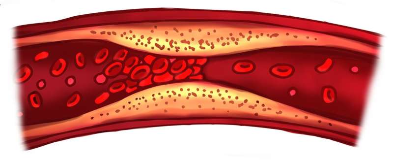
Thrombosis—clots forming inside blood vessels—can be very dangerous and can lead to heart attacks, strokes, and other serious health problems, the leading causes of death globally.
Professor Karlheinz Peter, a cardiologist and a researcher at the Baker Heart and Diabetes Institute and at the University of Melbourne, says that accurate and timely detection of thrombotic diseases is vital for improving patient outcomes, so that clinical interventions can quickly restore blood flow and oxygen supply to the affected organs and tissues.
However, the limitations of currently available diagnostic strategies often delay lifesaving treatment. "We are currently limited to using either indirect measurement of blood flow reduction in vessels, or functional readouts after the occurrence of potentially life-threatening tissue damage, such as following a heart attack," says Professor Peter.
A new preclinical study, led by Associate Professor Xiaowei Wang, Professor Peter and colleagues at the Baker Institute and published in the Arteriosclerosis, Thrombosis, and Vascular Biology journal, could ultimately provide a much-needed option to treat blood clots.
The researchers demonstrated successful targeting of Factor XIIa, a crucial component of the blood clot formation process. Wang and colleagues applied near-infrared fluorescence imaging, using a specific antibody that targets the activated form of Factor Xlla within the arteries of mice, to identify blood clots.
As Wang explains, molecular imaging uses markers that are specific to a particular disease, which makes the diagnosis more accurate and informative. This preclinical technology is gaining popularity, and the Baker Institute group has been studying it for several years.
Professor Peter says that while further investigation of the FXlla-specific antibody for thrombosis will be required in human studies, recently published work has found it to be a safe anticoagulant (without causing bleeding side-effects). As such, this antibody has great potential for both imaging and therapy.
The FXIIa-specific antibody was developed by CSL, one of the largest global biotech companies, with the Baker Institute providing the expertise in molecular imaging and atherothrombosis to test this promising reagent in the laboratory.
More information: Aidan P.G. Walsh et al, Activated Coagulation FXII (Factor XII): A Unique Target for In Vivo Molecular Imaging, Arteriosclerosis, Thrombosis, and Vascular Biology (2023). DOI: 10.1161/ATVBAHA.122.318883


