This article has been reviewed according to Science X's editorial process and policies. Editors have highlighted the following attributes while ensuring the content's credibility:
fact-checked
trusted source
proofread
Understanding dental pulp vitality—exploring clinical tests and histological analysis

Assessing the vitality of dental pulp is crucial in determining the appropriate treatment approach for dental conditions. Traditionally, clinicians have relied on subjective sensibility tests, such as temperature or electric stimuli, which provide indirect information and can be inconclusive.
These methods primarily evaluate neural responses, rather than the actual vascular circulation of the pulp. In recent years, innovative techniques like pulse oximetry and laser Doppler flowmetry have emerged as objective measures to assess pulp vitality. Additionally, histological analysis offers valuable insights into the morphology of dental pulp tissue.
This study, "Comparison of the vitality tests used in the dental clinical practice and histological analysis of the dental pulp," published in Bosnian Journal of Basic Medical Sciences, examines the correlation between clinical tests and histological analysis to gain a deeper understanding of dental pulp vitality.
The study was approved by the Slovenian national medical ethics committee, and it involved the examination of 26 healthy permanent premolars from seven patients aged 12 to 20 years. The teeth were free from any visible defects and scheduled for extraction due to orthodontic reasons. Clinical tests, including pulse oximetry for oxygen saturation measurement and an electric pulp tester, were conducted prior to the extractions.
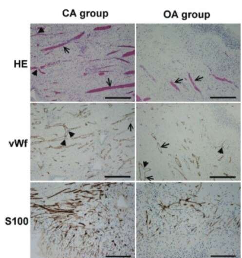
Histological analysis of the dental pulp tissue was performed using staining techniques and immunohistochemistry. The density of blood vessels and myelinated nerve fibers was evaluated, and statistical analyses were conducted to establish correlations between the clinical and histological findings.
The results of the study by Ana Tenyi, from the Department of Dental Diseases and Normal Dental Morphology, Medical Faculty University of Ljubljana, Slovenia and Aleksandra Milutinović, from the Institute of histology and embryology, Medical Faculty University of Ljubljana, Slovenia, found intriguing correlations between the clinical tests and histological analysis of dental pulp. First, a positive correlation was found between the volume density of blood vessels in the pulp tissue and oxygen saturation levels measured through pulse oximetry.
This finding, that a higher density of blood vessels is associated with increased oxygen saturation in the dental pulp, supports the validity of pulse oximetry as an objective method for assessing pulp vitality. Furthermore, a negative correlation was discovered between the volume density of myelinated nerve fibers and the lowest electrical voltage felt by the patients during the electric pulp testing.
This suggests that teeth with a higher density of nerve fibers have a lower threshold for electrical stimulation. Interestingly, teeth with closed apices exhibited a higher density of nerve fibers in the upper part of the dental pulp compared to teeth with open apices. However, there were no significant differences in the perception of electrical voltage between the two groups, indicating individual variations in sensitivity.
While the sample size and focus on healthy teeth for orthodontic extraction limit the generalizability of the findings, these results provide valuable insights into the relationship between clinical tests and histological characteristics of dental pulp. Pulse oximetry emerges as a reliable method for assessing pulp vitality, reflecting the vascularity and oxygen saturation of the pulp tissue.
The presence of a higher density of nerve fibers may contribute to a lower threshold for electrical stimulation, suggesting the involvement of neural factors in pulp sensibility. The positive correlation between blood vessel density and oxygen saturation, as well as the negative correlation between nerve fiber density and electrical voltage perception, provide valuable insights into the complex nature of dental pulp.
While further research with larger and more diverse samples is needed, these findings contribute to our understanding of dental pulp vitality and its relationship to clinical assessments. By demonstrating the utility of pulse oximetry in evaluating oxygen saturation and the importance of nerve fiber density in electrical sensibility, this study emphasizes the potential for more objective and accurate methods in assessing pulp vitality.
These findings have important implications for dental professionals. The use of pulse oximetry can provide a quantitative measurement of oxygen saturation in the dental pulp, enabling clinicians to assess the health and viability of the pulp tissue more accurately. This information can aid in determining the appropriate treatment approach, such as the need for pulp preservation or the possibility of pulp regeneration.
Moreover, the observed correlation between nerve fiber density and electrical sensibility highlights the influence of neural factors on pulp sensitivity. Understanding the role of nerve fibers in the perception of electrical stimuli can assist clinicians in evaluating and managing patient discomfort during dental procedures. It also emphasizes the importance of individual variations in sensitivity and the need for personalized treatment approaches.
The limitations of this study would be the small sample size and the focus on healthy teeth for orthodontic extraction. Further research with larger and more diverse samples is necessary to validate these findings and explore potential differences in pulp vitality among different dental conditions or age groups. Additionally, longitudinal studies could provide valuable insights into the changes in pulp vitality over time and the impact of aging on pulp health.
More information: Ana Tenyi et al, Comparison of the vitality tests used in the dental clinical practice and histological analysis of the dental pulp, Bosnian Journal of Basic Medical Sciences (2022). DOI: 10.17305/bjbms.2021.6841


