This article has been reviewed according to Science X's editorial process and policies. Editors have highlighted the following attributes while ensuring the content's credibility:
fact-checked
peer-reviewed publication
trusted source
proofread
Cell therapy that repairs cornea damage with patient's own stem cells achieves positive phase I trial results

A team led by researchers from Mass Eye and Ear, a member of Mass General Brigham, reports the results of a phase I trial of a revolutionary stem cell treatment called cultivated autologous limbal epithelial cell transplantation (CALEC), which was found to be safe and well-tolerated over the short term in four patients with significant chemical burns in one eye.
According to a study published August 18 in Science Advances, the patients who were followed for 12 months experienced restored cornea surfaces—two were able to undergo a corneal transplant and two reported significant improvements in vision without additional treatment.
While the phase I study was designed to determine preliminary safety and feasibility before advancing to a second phase of the trial, the researchers consider the early findings promising.
"Our early results suggest that CALEC might offer hope to patients who had been left with untreatable vision loss and pain associated with major cornea injuries," said principal investigator and lead study author Ula Jurkunas, MD, associate director of the Cornea Service at Mass Eye and Ear and an associate professor of ophthalmology at Harvard Medical School.
"Cornea specialists have been hindered by a lack of treatment options with a high safety profile to help our patients with chemical burns and injuries that render them unable to get an artificial cornea transplant. We are hopeful with further study, CALEC can one day fill this crucially needed treatment gap."
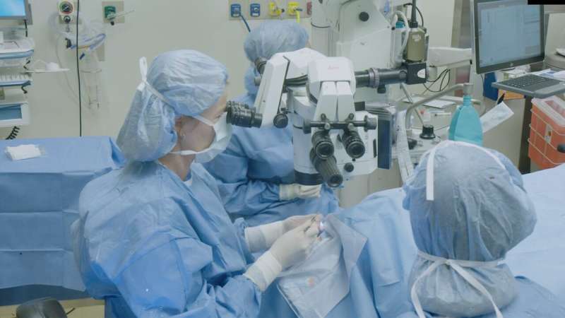
In CALEC, stem cells from a patient's healthy eye are removed via a small biopsy and then expanded and grown on a graft via an innovative manufacturing process at the Connell and O'Reilly Families Cell Manipulation Core Facility at Dana-Farber Cancer Institute. After two to three weeks, the CALEC graft is sent back to Mass Eye and Ear and transplanted into the eye with corneal damage.
The CALEC project is a collaboration between Jurkunas and colleagues in the Cornea Service at Mass Eye and Ear, researchers at Dana-Farber Cancer Institute, led by Jerome Ritz, MD, Boston Children's Hospital, led by Myriam Armant, Ph.D., and the JAEB Center for Health Research.
Expanding one's own stem cells to address limitations in existing treatments
People who experience chemical burns and other eye injuries may develop limbal stem cell deficiency, an irreversible loss of cells on the tissue surrounding the cornea. These patients experience permanent vision loss, pain and discomfort in the affected eye. Without limbal cells and a healthy eye surface, patients are unable to undergo artificial cornea transplants, the current standard of vision rehabilitation.
Existing treatment strategies have limitations and associated risks the CALEC procedure aims to address through its unique approach of using a small amount of a patient's own stem cells that can then be grown and expanded to create a sheet of cells that serves as a surface for normal tissue to grow back.
According to the authors, despite landmark studies describing an autologous stem cell approach over the past 25 years and similar methods being utilized in Europe, no U.S. research team had successfully developed a manufacturing process and quality control tests that met U.S. Food and Drug Administration (FDA) requirements or showed any clinical benefit.
"It was challenging to develop a process for creating limbal stem cell grafts that would meet the FDA's strict regulatory requirements for tissue engineering," said Ritz, executive director of the Connell and O'Reilly Families Cell Manipulation Core Facility at Dana-Farber and professor of medicine at Harvard Medical School. "Having developed and implemented this process, it was very gratifying to see encouraging clinical outcomes in the first cohort of patients enrolled on this clinical trial."
Studies like this show the promise of cell therapy for treating incurable conditions. Mass General Brigham's Gene and Cell Therapy Institute is helping to translate scientific discoveries made by researchers into first-in-human clinical trials and, ultimately, life-changing treatments for patients. The Institute's multidisciplinary approach sets it apart from others in the space, helping researchers to rapidly advance new therapies and pushing the technological and clinical boundaries of this new frontier.
Case studies hold early promise as clinical trial advances
In the phase I study, five patients with chemical burns to one eye were enrolled and biopsied. Four received CALEC; a series of quality control tests determined the cells in the fifth patient were unable to adequately expand. The CALEC patients were tracked for 12 months.
The first patient treated, a 46-year-old male, experienced a resolution of his eye surface defect, which primed him to undergo an artificial cornea transplant for vision rehabilitation. The second, a 31-year-old male, experienced a complete resolution of symptoms with vision improving from 20/40 to 20/30.
The third, a 36-year-old male, had his corneal defect resolved and his vision improved from hand motion—only being able to see broad movements like waving—to 20/30 vision.
The fourth, a 52-year-old male, initially did not have a successful biopsy that resulted in a viable stem cell graft. After re-attempting CALEC three years later, he underwent a successful transplant and his vision improved from hand motion to being able to count fingers. He then received an artificial cornea.
The researchers are finalizing the next phase of the clinical trial in 15 CALEC patients they are tracking for 18 months to better determine the procedure's overall efficacy. Their hope is that CALEC can one day become a treatment option for patients who previously had to endure long-term deficits when existing treatment options were not an option given the severity of their injuries.
More information: Ula Jurkunas, Cultivated autologous limbal epithelial cell (CALEC) transplantation: development of manufacturing process and clinical evaluation of feasibility and safety, Science Advances (2023). DOI: 10.1126/sciadv.adg6470. www.science.org/doi/10.1126/sciadv.adg6470


