This article has been reviewed according to Science X's editorial process and policies. Editors have highlighted the following attributes while ensuring the content's credibility:

fact-checked

peer-reviewed publication

trusted source

proofread

Menopausal hormone changes linked to cognitive deficits
Menopausal hormone changes linked to cognitive deficits
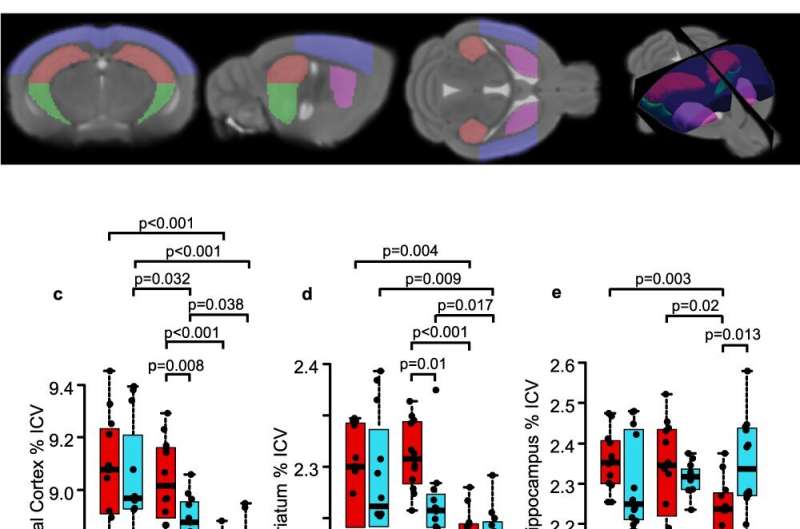
Female mice show abrupt brain substructure volume loss after midlife compared to males, and ovariectomized females have smaller dorsal hippocampal volumes than sham treated females at midlife. a Schematic showing timing of gonadectomy (GDX) or sham surgery, cognitive assessment by Morris Water Maze (MWM), in vivo MRI, and pathology (PATH) at young, midlife, and old ages. b In vivo MRI was collected from young, midlife, and old female and male mice. Substructure volumes visualized on the mean template (dorsal hippocampus = red, ventral hippocampus = green, cortex = blue, striatum = purple). Sham-treated female (red) and male (blue) volumes are expressed as a percentage of intercranial volume (ICV) for (c) frontal cortex, (d) striatum, and (e) dorsal hippocampus over the lifespan. Males showed gradual atrophy in frontal cortex and striatum from young to midlife to old age. In contrast, females maintained volumes through midlife, followed by atrophy from midlife to old age. Dorsal hippocampus showed atrophy in females from midlife to old age, while males did not have atrophy. Two-way ANOVA indicated a significant interaction between sex and age in dorsal hippocampus (p = 0.0059). n = 12 for all groups (c–e). p values were calculated by two-sided Welch’s t-test. Female sham-treated (solid) and GDX (diagonal lines) substructure volumes, assessed by MRI, expressed as a percentage of intercranial volume (ICV) are shown for (f) whole hippocampus, (g) dorsal hippocampus, and (h) ventral hippocampus. GDX females showed smaller dorsal hippocampus than sham females at midlife (p = 0.014). Female midlife sham n = 6 and midlife GDX n = 8, old sham n = 5 and old GDX n = 8. p values were calculated by two-sided Welch’s t-test. All box plots with center lines showing the medians, boxes indicating the interquartile range, and whiskers indicating a maximum of 1.5 times the interquartile range beyond the box. Credit: Nature Communications (2023). DOI: 10.1038/s41467-023-41723-7

A new study led by UCLA neurologist Dr. Rhonda Voskuhl sheds light on the underlying mechanisms linking menopause to cognitive deficits and brain atrophy, revealing a crucial role for estrogen receptor beta (ERβ) in astrocytes. The study, conducted on female mice, identified the specific brain regions and mechanisms responsible for the cognitive changes experienced during menopause.

The work is published in the journal Nature Communications.

The research found that loss of ovarian hormones in female mice during , but not at a younger age, induced cognitive impairment. This revealed that both aging and loss of estrogen were critical to cognitive deficits. Additionally, brain MRIs of these midlife female mice demonstrated atrophy of the dorsal hippocampus, a brain region central to memory and learning, and pathology revealed activation of astrocytes and microglia, with synaptic loss.

Selective deletion of estrogen receptor beta (ERβ) in astrocytes, a supportive brain cell, had the same detrimental effects on the brain as hormone loss, suggesting that ERβ in astrocytes plays a pivotal role in maintaining hippocampal function during menopause. To translate their findings to humans, the researchers showed that changes in in the hippocampus of estrogen deficient midlife involved abnormal glucose utilization, and expression of a key gene in this pathway also occurred in post-menopausal women.

Aiming to prevent deleterious effects of estrogen deficiency at midlife, mice treated with an ERβ ligand had improved cognition and reversal of the neuropathological changes observed in the dorsal hippocampus. While further research is needed to translate these findings into clinical applications for , the study marks a significant step toward understanding the brain's response to hormonal changes during menopause and offers hope for potential treatments in the future.

More information: Noriko Itoh et al, Estrogen receptor beta in astrocytes modulates cognitive function in mid-age female mice, Nature Communications (2023). DOI: 10.1038/s41467-023-41723-7

Journal information: Nature Communications
Citation: Menopausal hormone changes linked to cognitive deficits (2023, September 28) retrieved 28 April 2024 from https://medicalxpress.com/news/2023-09-menopausal-hormone-linked-cognitive-deficits.html
This document is subject to copyright. Apart from any fair dealing for the purpose of private study or research, no part may be reproduced without the written permission. The content is provided for information purposes only.

Explore further

Importance of dietary changes and physical activity to mitigate weight gain during midlife

62 shares

Feedback to editors