This article has been reviewed according to Science X's editorial process and policies. Editors have highlighted the following attributes while ensuring the content's credibility:
fact-checked
trusted source
proofread
Enhanced osteochondral repair with hyaline cartilage formation using an extracellular matrix-inspired natural scaffold

Osteochondral defects, often caused by injury or various pathologies, typically lead to the onset of osteoarthritis, and may ultimately result in the comprehensive degradation of the joint and consequent disability. Despite a range of clinical treatments available, such as microfracture, autologous cell transplantation, and osteochondral autografts, persistent issues like inadequate regeneration and graft failure continue to pose substantial challenges for osteochondral repair.
In particular, because of the avascular and limited regenerative properties of osteochondral tissue, inappropriate repair can generate fibrous cartilage and subchondral bone, causing the degeneration of osteochondral tissue and eventually leading to repair failure. Thus, biomimetic tissue engineering strategies were developed to treat osteochondral defects by combining substitute materials. Nevertheless, the challenge of regenerating osteochondral tissue persists, primarily due to the tissue lineage differences and the anisotropic physiological characteristics.
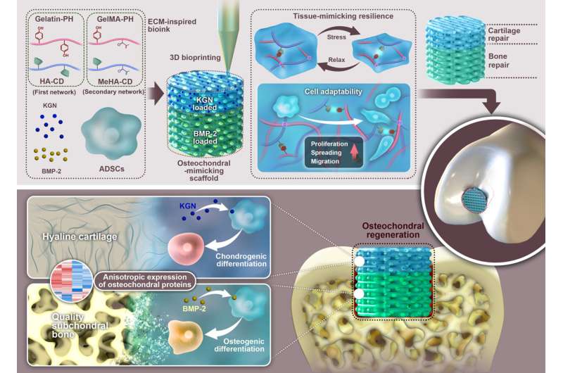
Taking inspiration from the chemical elements inherent in the natural ECM, a study published in Science Bulletin designed a novel bioink composed of natural polysaccharides and polypeptides to fabricate an ECM-mimicking scaffold for osteochondral regeneration. Consisting of filamentous substances such as polysaccharides and polypeptides, the natural ECM forms a hydrogel-like structure with a hierarchical arrangement. It provides essential mechanical and structural reinforcement for cell survival and phenotype modulation.
To create an ECM-mimicking microenvironment for cellular support, polysaccharides, such as hyaluronic acid (HA) and chitosan, or polypeptides, such as gelatin and silk fibroin, were used to make scaffolds. However, as the selected polypeptides and polysaccharides only have one function each, current hydrogels that attempt to mimic the ECM face difficulties in replicating the structure and biological function of the natural ECM.
In this study, researchers hypothesized that biomimetically fabricated scaffolds based on polypeptides and polysaccharides would offer a unique approach for encouraging cell survival, differentiation, and osteochondral regeneration. To put this concept into practice, the natural polysaccharide HA and polypeptide gelatin were meticulously modified to form a novel bioink (MeGH) with reversible guest-host and rigid covalent networks.
Through the formed guest-host and covalent networks between the HA and gelatin chains, the resultant MeGH not only exhibited outstanding biocompatibility, cell adaptability, and biodegradability but also had excellent mechanical properties to cater to the environment of load-bearing osteochondral tissue.
Furthermore, benefiting from the drug-loading group of the bioink, osteogenic and chondrogenic drugs could be precisely integrated into the specific zone of the scaffold, endowing it with an osteochondral-specific microenvironment to facilitate cartilage and bone differentiation. In the case of rabbits with osteochondral defects, the ECM-inspired scaffold successfully promoted the development of hyaline cartilage and robust subchondral bone in a manner that imitates the natural gradual osteochondral transition.
In summary, inspired by the inherent chemical components of natural ECM, this study first designed and synthesized a natural hydrogel scaffold that mimics extracellular matrix. This scaffold enhances cell survival and provides a tissue-specific microenvironment to facilitate bone and cartilage differentiation, thereby accelerating osteochondral repair and the formation of hyaline cartilage. This study provides new insights for the clinical repair of osteochondral defects.
This study was led by Prof. Yingfang Ao, Prof. Jianquan Wang and Dr. Xiaoqing Hu (Beijing Key Laboratory of Sports Injuries, Institute of Sports Medicine of Peking University, Peking University Third Hospital).
More information: Wenli Dai et al, Enhanced osteochondral repair with hyaline cartilage formation using an extracellular matrix-inspired natural scaffold, Science Bulletin (2023). DOI: 10.1016/j.scib.2023.07.050


