This article has been reviewed according to Science X's editorial process and policies. Editors have highlighted the following attributes while ensuring the content's credibility:
fact-checked
proofread
New insights into heterotopic ossification: Progenitor cells play a key role in aberrant bone formation

In a new study published in Bone Research, a team of researchers from Johns Hopkins University and colleagues, using a combination of lineage tracing and single-cell RNA sequencing (scRNA-seq), has delved into the contribution of synovial/tendon sheath progenitor cells to heterotopic bone formation.
The researchers identified a distinct population of Tppp3+ tendon progenitor cells that actively participated in the formation of ectopic bone in vivo. Their findings provide new insights into the intricate cellular processes driving heterotopic ossification and offer potential therapeutic targets to prevent and treat this pathological condition.
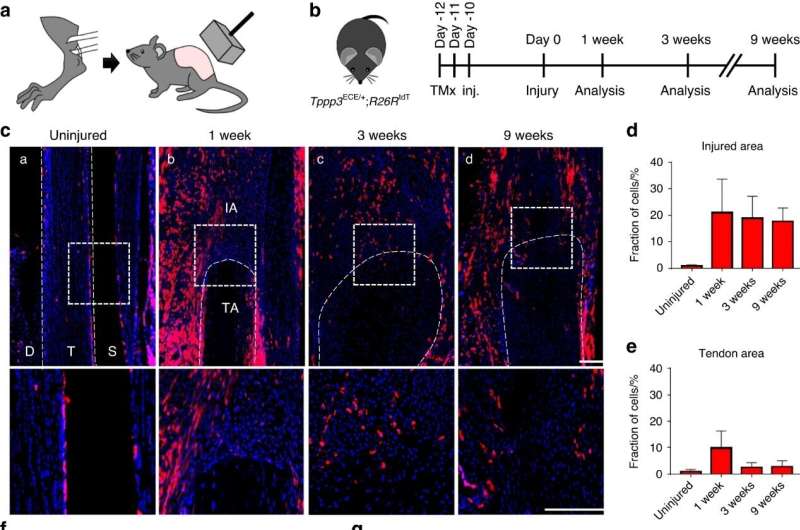
The study revealed that Tppp3+ progenitor cells, primarily located in the tendon sheath, rapidly expanded in response to HO induction, significantly contributing to the formation of heterotopic cartilage and bone in both tendon and joint-associated HO.
Transcriptomic analysis indicated that Tppp3+ cells possess primitive progenitor properties and may regulate HO formation by releasing soluble molecules that promote osteogenic differentiation. Under normal conditions, Tppp3-tdT+ cells were mainly localized in the tendon sheath with minimal activity in the tendon body.
However, upon HO induction, their numbers rapidly increased at the injury site. Tppp3 was predominantly expressed in mesenchymal cells, indicating a progenitor cell phenotype. Furthermore, scRNA-seq analysis revealed Tppp3 as an early progenitor cell marker for either tendon or osteochondral cells, offering insights into the dynamic behavior of Tppp3+ progenitor cells during injury.
These cells were found to contribute significantly to heterotopic cartilage formation during the endochondral ossification phase, with around one-fifth of cartilage cells derived from Tppp3+ progenitors. Additionally, Tppp3+ cells played a crucial role in the osseous phase of heterotopic ossification in the Achilles tendon, contributing to the osteoblastic lineage at 9 weeks after injury.
Notably, they also participated in tendon remodeling and the formation of a tendon-like matrix during the repair process. In the hip postarthroplasty HO model, Tppp3+ cells contributed to the formation of heterotopic cartilage in the synovium and expanded in the synovium and periosteum, acquiring an osteoprogenitor phenotype and contributing to ectopic bone formation.
The presence and contribution of Tppp3+ cells in human pathological samples were confirmed, highlighting the relevance of these findings to human cases of HO.
The research contributes significantly to the field of regenerative medicine and sheds light on the regulatory role of Tppp3+ progenitor cells in HO formation and provides valuable insights for potential therapeutic strategies to target this pathological process. The identification of this newly defined population of tendon progenitors that contribute to ectopic bone formation may pave the way for the development of novel therapeutic approaches to prevent and treat HO.
More information: Ji-Hye Yea et al, Tppp3+ synovial/tendon sheath progenitor cells contribute to heterotopic bone after trauma, Bone Research (2023). DOI: 10.1038/s41413-023-00272-x


