This article has been reviewed according to Science X's editorial process and policies. Editors have highlighted the following attributes while ensuring the content's credibility:
fact-checked
peer-reviewed publication
trusted source
proofread
First digital atlas of human fetal brain development published

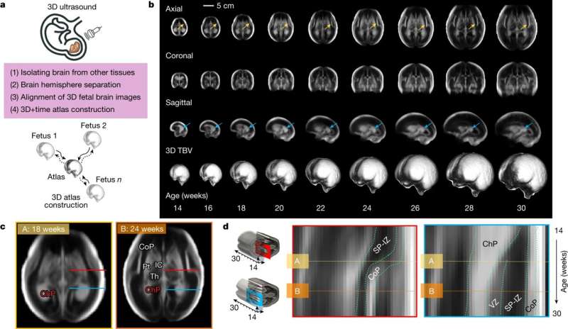
A team of more than 200 researchers around the world, involving multiple health and scientific institutions, led by the University of Oxford, has published the first digital atlas showing the dynamics of normative maturation of each hemisphere of the fetal brain between 14 and 31 weeks' gestation—a critical period of human development. The article is titled "Normative spatiotemporal fetal brain maturation with satisfactory development at 2 years" and appears in the journal Nature
The atlas was produced using more than 2,500 3-dimensional ultrasound (3D US) brain scans that were acquired serially during pregnancy from 2,194 fetuses in the INTERGROWTH-21st Project, which is a large population-based study of healthy pregnant women living in eight diverse geographical regions of the world (including five in the Global South), whose children had satisfactory growth and neurodevelopment at 2 years of age.
The study is unique because, for the first time, an international dataset of 3D US scans, collected using standardized methods and equipment, has been analyzed with advanced artificial intelligence (AI) and image processing tools to construct a map showing how the fetal brain matures as pregnancy advances.
Demonstrating remarkably similar patterns of fetal brain growth and development across diverse populations represents an important scientific advance in the field of neuroscience. The results are entirely consistent with previously reported findings, from the same INTERGROWTH-21st population, for fetal skeletal growth, newborn size and infant neurocognitive development. The results also highlight a vitally important public health message: a mother's health, educational, nutritional and environmental needs must be met to ensure that her child's body and brain develop healthily.
The findings add to the global impact of the INTERGROWTH-21st Project, which has previously produced international standards for fetal growth, newborn size and the postnatal growth of preterm babies, that are being widely adopted across the world for clinical and research purposes.
Professor Ana Namburete, the first author, whose research group developed the machine learning methods, said, "Using AI we enhanced the visibility of brain structures in the 3D US images, and generated an average depiction of the brain at each week of pregnancy during a critical period of development. Uniquely, our atlas captured patterns of brain growth from as early as 14 weeks' gestation—filling a 6-week knowledge gap in our understanding of early fetal brain maturation.
"We also revealed significant asymmetries in brain maturation: for example, in the region associated with language development, which peaked at 20–26 weeks' gestation and persisted thereafter without any differences between the sexes."
Professor Stephen Kennedy, co-Principal Investigator of the INTERGROWTH-21st Project, who jointly led the study, said, "The atlas will help scientists answer complex biological questions about the fetal origins of cognitive function in childhood, such as how language is acquired. Using the atlas in combination with the soon to be published international standards describing the complementary growth of the fetal brain will be a valuable clinical tool in specialized, referral centers when brain development appears abnormal on ultrasound."
Professor José Villar, co-Principal Investigator of the INTERGROWTH-21st Project, who jointly led the study, said, "This is the latest step in the systematic study of early human growth and development that confirms, using the most advanced research methodology applied to a large number of fetal brain scans, the similarities of growth and development of humans across the world when health, educational, nutritional and environmental needs are met: humans are very similar in all domains, including their brains, when conditions are adequate."
The fetal brain atlas can be found freely available online.
More information: Ana I. L. Namburete et al, Normative spatiotemporal fetal brain maturation with satisfactory development at 2 years, Nature (2023). DOI: 10.1038/s41586-023-06630-3


