This article has been reviewed according to Science X's editorial process and policies. Editors have highlighted the following attributes while ensuring the content's credibility:

fact-checked

trusted source

proofread

Noninvasive technique helps visualize inflammatory cells in human heart
Noninvasive technique helps visualize inflammatory cells in human heart
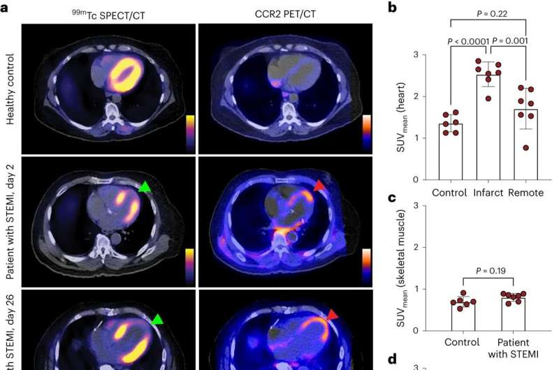
CCR2 imaging in healthy controls and individuals after STEMI. a, Representative 99mTc tetrofosmin (99mTc) SPECT/CT and differential 64Cu-DOTA-ECL1i (CCR2) PET/CT fused images of healthy controls and patients with STEMI. SPECT/CT perfusion and CCR2 PET/CT images were coregistered and comparative anatomic slices are displayed. Differential 64Cu-DOTA-ECL1i images were corrected for blood activity. The green and red arrows denote the infarct region. The color scale bar indicates normalized relative tracer uptake. b, 64Cu-DOTA-ECL1i myocardial signal. For patients with STEMI, differential SUVmean was calculated in the infarct and remote regions. Statistical significance was determined using a one-way analysis of variance (ANOVA). Controls, n = 6; patients with STEMI, n = 7. Control versus infarct: P = 0.00008; infarct versus remote: P = 0.001; control versus remote: P = 0.22). c, 64Cu-DOTA-ECL1i skeletal muscle signal. Statistical significance was determined using a two-sided Mann–Whitney U-test. Controls, n = 6, patients with STEMI, n = 7. d, 64Cu-DOTA-ECL1i myocardial signal as a function of time after STEMI. Each data point represents an individual patient. The mean value for controls with s.d. is displayed as a line with gray zones. Patients with STEMI, n = 7. e, Corresponding 17-segment model polar maps of SPECT/CT perfusion and myocardial wall motion and CCR2 PET/CT from a representative individual. 99mTc tetrofosmin SPECT/CT and wall motion polar maps are displayed as the percentage of maximal value (100%). The CCR2 PET/CT polar map represents SUVmean values. f, Linear regression analysis examining the association between CCR2 tracer uptake and wall motion score. P = 0.03 and r2 = 0.4; n = 7. Each data point represents an individual patient. The error bars represent the s.d. Credit: Nature Cardiovascular Research (2023). DOI: 10.1038/s44161-023-00335-6

A study in Nature Cardiovascular Research by researchers at Washington University School of Medicine in St. Louis explores a new, noninvasive imaging technique that helps scientists visualize immune cells in the human heart.

Inflammatory contribute to , increasing the risk of a heart attack and heart failure. However, technologies to identify the immune cell types responsible for inflammation in the muscular tissue of the heart—the myocardium—are limited. The researchers developed a radiotracer to help visualize a subset of inflammatory immune cells marked by the expression of C-C chemokine receptor 2, which includes harmful populations of monocytes and macrophages.

The research was performed by Yongjian Liu, Ph.D., a professor of radiology at Mallinckrodt Institute of Radiology (MIR), and Kory J. Lavine, MD, Ph.D., an associate professor of medicine, of , and of pathology and immunology, in collaboration with colleagues from the PET Radiotracer Translation and Resource Center and Immuno-Fib HF Network.

The research team established the feasibility of noninvasively visualizing inflammatory immune cells using in patients who had suffered heart attacks. Their findings may help identify individuals who may benefit from immunomodulatory therapies.

The noninvasive imaging technology developed by the research team "could not only identify individuals best suited for immunomodulatory therapies, but also assess treatment outcomes for better patient management," concluded the authors, pointing to the potential value for enriching clinical trials and providing precision medicine as the technology is developed further. Future studies will focus on the expansion of the patient population and evaluate prognostic implications of the imaging on cardiac remodeling, function and clinical outcomes.

More information: Kory J. Lavine et al, CCR2 imaging in human ST-segment elevation myocardial infarction, Nature Cardiovascular Research (2023). DOI: 10.1038/s44161-023-00335-6

Citation: Noninvasive technique helps visualize inflammatory cells in human heart (2023, October 24) retrieved 30 April 2024 from https://medicalxpress.com/news/2023-10-noninvasive-technique-visualize-inflammatory-cells.html
This document is subject to copyright. Apart from any fair dealing for the purpose of private study or research, no part may be reproduced without the written permission. The content is provided for information purposes only.

Explore further

Researchers find subset of cardiac fibroblasts involved in cardiac inflammation under stress

4 shares

Feedback to editors