This article has been reviewed according to Science X's editorial process and policies. Editors have highlighted the following attributes while ensuring the content's credibility:
fact-checked
peer-reviewed publication
trusted source
proofread
New study reveals how HIV binds to our T cells

A new study reveals for the first time the steps through which human immunodeficiency virus (HIV) binds to the receptors on the membranes of T cells—white blood cells that fight infection. The finding could have implications for developing new therapies.
There is currently no vaccine or cure for HIV. Antiretroviral therapy (ART) helps people living with HIV suppress the virus to undetectable levels, but those patients must take these medications for the rest of their lives, and for some, the drugs' effectiveness fades over time. Scientists know that HIV infects a host by first binding to a cell-surface receptor called CD4.
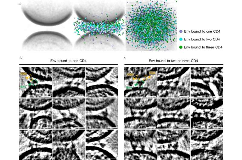
Using an imaging technique known as cryogenic electron tomography (cryo-ET), a team was able to visualize, for the first time, how HIV-1, the most common type of HIV, interacted with virus-like particles (VLP) carrying CD4 receptors, mimicking how HIV interacts with T cells in nature. The new work, published November 22 in Nature, uncovered the stepwise interactions between the proteins of the HIV-1 and VLP membranes, including the structures of the intermediate stages as HIV-1 binds to a host.
"Our study shows the very early stages of how this terrible disease begins and the steps of how it engages with receptors," to then fuse membranes with T cells, says Walther Mothes, Ph.D., Paul B. Beeson Professor of Medicine at Yale School of Medicine and principal investigator. The team hopes its findings will lead to new inhibitory HIV medications that specifically target these HIV conformations.
Cryo-ET reveals how HIV-1 binds to CD4 receptor
The Mothes Laboratory investigates how viruses spread and cause disease, from animal models to single molecules. In the most recent study, the team used murine leukemia virus (MLV) to produce the VLPs with CD4 receptors. Then, they observed a mixture of HIV-1 and VLPs using cryo-ET and studied the interactions that occurred on the membranes.
The researchers saw that the HIV-1 and VLP gathered in small clusters and formed rings. When the membranes were farther apart, HIV-1 was bound to only one CD4. As the membranes moved closer together, HIV-1 bound to a second and third CD4. "We believe these three intermediate steps represent how HIV naturally binds to CD4 on T cells," says Mothes.
Their findings support a related paper, also in Nature, published by the California Institute of Technology's Bjorkman Lab, led by Pamela Bjorkman, Ph.D., David Baltimore Professor of Biology and Biological Engineering. Bjorkman's group had engineered atomic models of the conformational states of HIV as it binds to one or two CD4 receptor molecules. "But they didn't know if their models existed in nature," says Mothes. "Our studies on real membranes show that they do."
Therapeutic implications for HIV and COVID-19
By creating inhibitors that target the intermediate HIV conformations, scientists hope to be able to intervene before HIV infects a host cell. "We have a window where we can specifically target these conformational states with antibodies and drugs," says Mothes.
One goal is to stop HIV, while not interfering with other molecules that are beneficial to cells.
"Imagine HIV viruses as rogue cars running on roads. Current drugs block the lanes to stop virus spread, but they also affect other cars in the traffic," explains Wenwei Li, Ph.D., associate research scientist in the Mothes Laboratory and first author of the study. "We are learning what the viruses look like—the color, size, and shape—so that we can specifically target them with drugs, pulling over the viruses without affecting the other cars in the traffic."
After a virus binds to a host, the membranes fuse together, allowing the virus to proliferate. In future studies, the team wants to study this fusion. "There are two steps to infection. We observed step one in this study," says Li. "And now we are looking for step two."
The study also could have implications beyond HIV. The team plans to apply its techniques to better understand SARS-CoV-2 infection, which could lead to the development of better drugs for COVID-19.
More information: Wenwei Li et al, HIV-1 Env trimers asymmetrically engage CD4 receptors in membranes, Nature (2023). DOI: 10.1038/s41586-023-06762-6


