This article has been reviewed according to Science X's editorial process and policies. Editors have highlighted the following attributes while ensuring the content's credibility:
fact-checked
trusted source
proofread
Heart organoids simulate pregestational diabetes-induced congenital heart disease

An advanced human heart organoid system can be used to model embryonic heart development under pregestational diabetes-like conditions, researchers report in the journal Stem Cell Reports.
The organoids recapitulate hallmarks of pregestational diabetes-induced congenital heart disease found in mice and humans. The findings also showed that endoplasmic reticulum (ER) stress and lipid imbalance are critical factors contributing to these disorders, which could be ameliorated with exposure to omega-3s.
"The new stem cell-based organoid technology employed will enable physiologically relevant studies in humans, allowing us to bypass animal models and obtain more information about relevant disease mechanisms, accelerating drug discovery and medical translation," says senior study author Aitor Aguirre of Michigan State University.
Congenital heart disease is the most common type of congenital defect in humans. Pregestational diabetes—diabetes affecting the mother before and during the first trimester of pregnancy—is an important factor contributing to congenital heart disease and is present in a significant, growing population of diabetic female patients of reproductive age.
Newborns from mothers with pregestational diabetes can have up to a 12-fold increased risk of congenital heart disease. Unfortunately, pregestational diabetes is hard to manage clinically due to the sensitivity of the developing embryo to glucose oscillations, and it represents a critical health problem for the mother and the fetus.
The limited access to human tissues for research of early-stage disease has resulted in an overreliance on animal models. But it remains unclear to what extent rodent models recapitulate abnormalities present in humans, given critical species differences in heart size, cardiac physiology, electrophysiology, and bioenergetics.
In addition, rodent models and many in vitro cell models rely on aggressive diabetic conditions, leading to exaggerated features that may not be clinically relevant.
"Advances in biotechnology and bioengineering are enabling the creation of human mini-organs in vitro," Aguirre says. "These mini-organs can currently be used to understand human disease much better without the drawbacks of animal models."
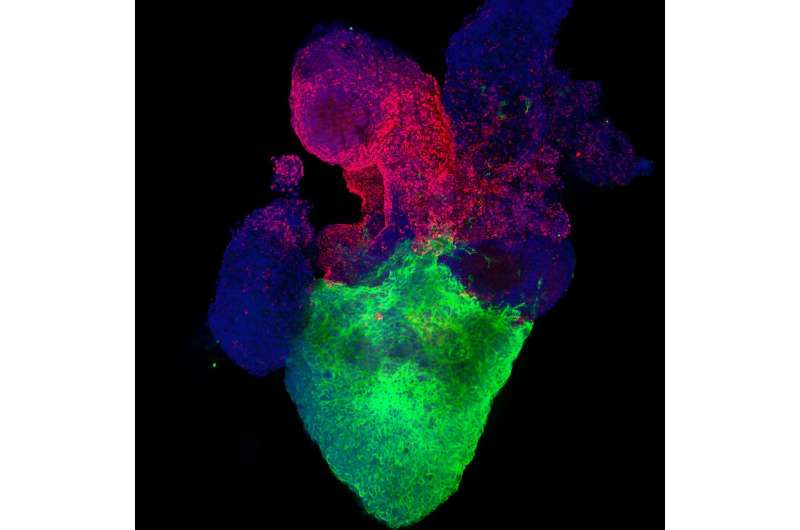
In the new study, Aguirre and his team used an advanced heart organoid model derived from human pluripotent stem cells. This model recapitulates human heart development during the first trimester, including critical steps such as chamber formation, vascularization, cardiac tissue organization, and relevant cardiac cell types.
To specifically model the effects of pregestational diabetes, the researchers modified culture conditions to accurately reflect reported physiological levels of glucose and insulin in patients.
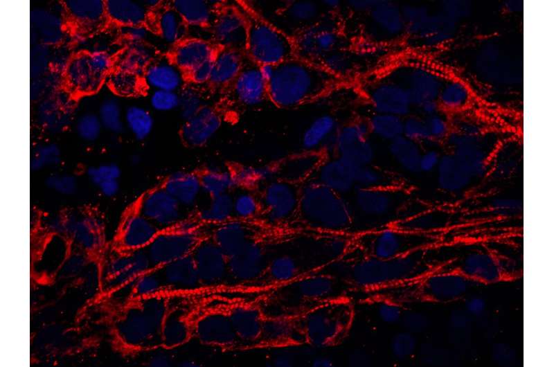
The resulting pregestational diabetes heart organoids (PGDHOs) developed features observed in previous mouse and human studies. For example, the diabetic human heart organoids were larger, suggesting signs of cardiac hypertrophy—a first hallmark of maternal pregestational diabetes. This observation was confirmed by studying cardiomyocyte size.
The PGDHOs also displayed arrhythmias and a reduction in beat frequency, which has been observed in neonatal rats from diabetic mothers. In addition, single-cell transcriptomic analysis of PGDHOs revealed a reduction in cardiomyocyte numbers, a significant expansion of tissue on the outer surface of the heart, and the absence of a well-developed vasculature at early developmental stages.

The PGDHOs also exhibited increased accumulation of reactive oxygen species (ROS), revealing increased oxidative stress and mitochondrial swelling—also hallmarks of diabetic embryonic heart conditions.
A significant portion of ROS was localized to the ER and could be impairing its function, leading to a condition known as ER stress. Moreover, the PGDHOs revealed a clear imbalance in very long chain fatty acids, particularly affecting omega-3 polyunsaturated fatty acids, which are mostly synthesized in the ER.
Taken together, these results point to a major ER-induced lipid imbalance in PGDHOs. This imbalance is linked to the degradation of fatty acid desaturase 2 (FADS2)—a key lipid biosynthesis enzyme presents in the ER—by the IRE1-dependent mRNA decay (RIDD) pathway, which has been implicated in several other cardiac conditions.
In an attempt to remedy the effects of ER stress, the researchers tested several potentially therapeutic compounds on PGDHOs. A mixture of omega-3 fatty acids ameliorated diabetic features, while targeting inositol-requiring enzyme 1 (IRE1) reduced cardiomyocyte hypertrophy. All the compounds also restored FADS2 levels.
"The organoids still lack some features that could be important, such as external vascularization and outflow tract, and better chamber formation, so we might still be missing important aspects of congenital heart disease and diabetic cardiomyopathy," Aguirre says.
"On one hand, we want to partner with clinicians to establish the efficacy and safety of our findings in pregnant women. On the other hand, we want to apply our organoid model to other conditions affecting congenital heart disease so we can improve the lives of these children in the future."
More information: ER stress and lipid imbalance drive diabetic embryonic cardiomyopathy in an organoid model of human heart development, Stem Cell Reports (2024). DOI: 10.1016/j.stemcr.2024.01.003. www.cell.com/stem-cell-reports … 2213-6711(24)00006-7


