This article has been reviewed according to Science X's editorial process and policies. Editors have highlighted the following attributes while ensuring the content's credibility:
fact-checked
peer-reviewed publication
trusted source
proofread
Researchers develop 3D model to better treat neurological disorders

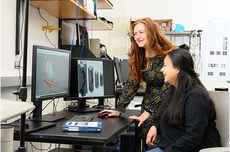
A 3D model developed by West Virginia University neuroscientists shows how implantable stimulators—the kind used to treat chronic pain—can target neurons that control specific muscles to provide rehabilitation for people with neurological disorders such as stroke and spinal cord injuries.
The study, including the model, was published Communications Biology.
The device, implanted on or near the spinal cord, works by delivering an electrical signal through a thin wire. To treat paralysis, stimulation targets specific parts of the spinal cord to help restore muscle function and movement. However, the effectiveness of the device has been constrained due to insufficient understanding of where motoneurons that connect to specific muscles are situated within the spinal cord.
"If we really want to maximize the usefulness of these implants, we want to be able to select specific motoneurons that would activate specific muscles and assist with the movement in the right way and at the right time," said Valeriya Gritsenko, associate professor in the WVU School of Medicine, departments of Human Performance—Physical Therapy, Neuroscience and the Rockefeller Neuroscience Institute. "Scientists want to use a model to figure out where to implant this system."
Gritsenko plans to lead a team in building more sophisticated models of the neuromuscular system.
With further studies and testing, researchers are hopeful to gain a better understanding of the extent to which these devices can improve muscle function.
To conduct the study, researchers first created a 3D model of the motoneuron locations in the macaque—an Old World monkey—spinal cord and compared it to the current knowledge of the human spinal cord. They also created 3D models of the musculoskeletal anatomy of the macaque and human right upper limb and compared those.
"We were looking at the differences and changes in muscle lengths across different postures in both the human model and the monkey model," said Rachel Taitano, a doctoral student in medicine and neuroscience from Fairfax, Virginia, and lead author of the study. "The musculoskeletal model of the monkey shows that the biomechanics are similar to humans even though the species have differences in the muscles they use, the muscles they have, and different orientations and functionality."
The study shows a close match in the distribution, or depth, of motoneuron pools along the spinal cord in macaques and humans. Those findings will allow scientists to gain precision in delivering treatment.
"Some motoneuron pools are deeper inside the spinal cord and others are closer to the surface," Gritsenko explained. "This model allows us to look in depth to where those motoneuron pools might be closest to the surface. That's where you would want to stimulate to potentially activate those muscles."
Gritsenko, who served as the primary investigator, explained that "knowing the spinal organization of motoneuron pools—groups of cells that connect to a single muscle—can reveal something fascinating. Our complex musculoskeletal system has evolved over time to enable the wide range of movements we see in all primates, including us humans. The team found that our spinal cords have built-in 'maps' that reflect this complex function. This 'map' helps simplify the control of our complex body by the spinal cord. It is like having an autopilot right inside the spine."
Another colleague on the project, Sergiy Yakovenko, associate professor in the WVU School of Medicine, departments of Human Performance—Exercise Physiology, Neuroscience and RNI, has conducted similar studies on the spinal cord anatomy in quadrupedal animals. The new findings show how well the anatomy of the spinal cord is conserved across animals and how closely it reflects the actions of the muscles.
Results from an applied science study that can be used to benefit patients in a clinical setting is what Taitano said drew her to the project.
"I think we can get a lot of information from non-invasive studies," said Taitano, who holds an undergraduate degree in biomedical engineering. "Now that we can apply these findings on the millimeter and the nanometer scale, we can fabricate devices to apply what we're seeing in a model like this."
With the project complete, Taitano moves on to the medical degree portion of her program this summer.
"Rachel's background was very instrumental in making the study a success," Gritsenko said. "I would definitely like to see more of this kind of interdisciplinary collaboration with graduate students working on projects with colleagues from medical and engineering departments."
Gritsenko said scientists at two other universities have expressed interest in using the model to explore how the stimulation technology can be improved. She also plans to collaborate with a primate researcher at another university to validate the study's findings in animal models.
"We want to do a muscle stimulation test based on the model predictions and see if we get the expected results," she said. "We can try that first with monkeys and then, if it works out, we can try it with humans to further check that it is a good model for guiding these surgeries."
More information: Rachel I. Taitano et al, Muscle anatomy is reflected in the spatial organization of the spinal motoneuron pools, Communications Biology (2024). DOI: 10.1038/s42003-023-05742-w


