Radiology & Imaging
New technology can detect kidney diseases earlier than standard methods
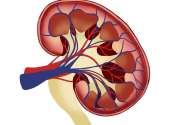
Using an advanced scanner, researchers from Aarhus University, among others, have developed a technology that can detect the earliest changes in the kidney when scar tissue begins to form. Their study is published in the ...
16 hours ago
0
0