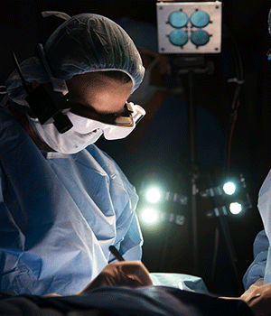
New imaging technique sharpens surgeons' vision

Which superhuman power would you choose for help on the job? For Dr. Julie Margenthaler, it's a technology that brings to mind X-ray vision, used for the first time Monday during an operation to remove a patient's lymph node.
"It's like I'm in a sci-fi movie," Margenthaler said after she put on goggles that allowed her to see the patient's lymph node light up with a fluorescent blue glow invisible to the naked eye. A video camera projected the surgeon's visual field onto the screen inside the goggles and on a computer screen in the operating room.
The new technology developed at Washington University may eventually be used to see microscopic cancer cells during surgery and enable a more thorough removal of tumors. For now, the research team is testing the goggles on 20 to 30 breast cancer and melanoma patients to find lymph nodes that will be tested for possible spread of the cancer. Karen Clodfelter, 67, of St. Louis, was the study's first patient.
"It was a lot less cumbersome than I thought it would be," Margenthaler said of the lightweight goggles that telescope the surgeon's visual field. "When I had (Clodfelter's) lymph node between my two forceps it was lighting up very well."
After the surgical staff helped Margenthaler put on the goggles, the team cheered when the process worked as planned.
"We're all interested in finding some way to perfect our surgical technique and ultimately find cancer cells earlier," Margenthaler said after the one-hour procedure at Barnes-Jewish Hospital.
The research team of surgeons and biomedical engineers has been working on the new imaging technique for several years. The idea came out of discussions among surgeons and scientists about the challenges of identifying the margins of tumor cells during cancer surgeries. Samuel Achilefu, a professor of radiology and biomedical engineering, remembered the night-vision goggles used in the Persian Gulf War and thought maybe a similar technology could be used in the operating room.
"The whole idea is to simplify the way we image patients and the way we guide surgery," said Achilefu, who leads the team. "Once you put on these goggles, you can detect tissues with high sensitivity. It gives the information processed in real time about the tumor's location, shape and about the tissue surrounding it so that you can make immediate medical decisions."
Currently, surgeons use X-ray or MRI images taken beforehand to plan the removal of tumors during surgery. But because the images don't pick up smaller clusters of tumor cells, surgeons take extra surrounding tissue and nearby lymph nodes to check for spread of the cancer. The tissue is sent to the lab, and if additional cancer is found, more surgery is scheduled. Up to one-fourth of patients who have breast lumpectomies have to return for a second surgery for leftover cancer cells.
During a cancer surgery, the suspicious cells aren't always distinguishable, said Dr. Ryan Fields, who specializes in melanoma surgery. Fields plans to use the goggles for the first time next month.
The researchers also developed a new dye formula that can target cancer cells to make them glow during surgeries using the goggles. The dye is awaiting approval for use in humans. In mouse studies, the system detected tumors as small as 1 millimeter in diameter, according to the team's research funded by the National Cancer Institute and published in the Journal of Biomedical Optics. The team has applied for a patent for the technology.
Achilefu hopes the technology can one day be used in telemedicine, by beaming the surgeon's vision to screens in medical school classrooms. Alternately, a surgeon using the goggles in another country or a rural area could send the images to St. Louis doctors to help guide the operation. The final, wireless version of the goggles is expected to be finished by the end of the year. The cost of the system should be under $10,000, he estimates.
"It's such a nice device that I think already we are getting requests from other parts of the world for prototypes," Achilefu said. "I can see it being used in so many places."
©2014 St. Louis Post-Dispatch
Distributed by MCT Information Services


