Distinctive developmental origin for a drainage tube in the eye
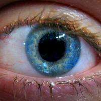
A Jackson Laboratory based research team has conducted a comprehensive exploration of an eye structure known as Schlemm's canal: a key gatekeeper for the proper flow of eye fluid, presenting a number of insights relevant to glaucoma and other diseases.
For the study publishing July 22 in the Open Access journal PLOS Biology, the researchers at JAX and Tufts University School of Medicine in Boston developed a new, "whole-mount," three-dimensional approach to analyse mouse models that have been engineered to host fluorescent proteins, to determine how Schlemm's canal forms in the eye and in relation to neighbouring tissues.
Due to its roles in fluid flow and intraocular pressure, Schlemm's canal is directly involved in glaucoma, a blinding disease that affects more than 70 million people worldwide.
The report, according to first author Krishnakumar Kizhatil, Ph.D., an associate research scientist in the laboratory of JAX Professor and Howard Hughes Medical Investigator Simon W.M. John, Ph.D., "provides new understanding and tools that will facilitate molecular understanding of Schlemm's canal and its critical—but poorly understood—roles in ocular physiology, immunity and health."
The researchers show that Schlemm's canal forms from blood vessels by a novel process of vascular development that they name canalogenesis. Canalogenesis has some similarities to previously established processes of vascular development—namely angiogenesis, vasculogenesis and lymphangiogenesis—but also has unique features that make it distinct from each of them. They also identify the first molecule to be functionally implicated in early Schlemm's canal development, namely the KDR receptor, which is also known to play a key role in blood vessel development.
Importantly, the research demonstrates that the endothelial cells lining this drainage tube (called SECs) are novel, having a blend of properties of both of blood and lymphatic endothelial cells. "Thus, Schlemm's canal is a unique vessel with endothelial cells that are highly specialized for its complex functions," Kizhatil says. "This resolves a long-standing controversy about the cellular phenotype of SECs."
Study coauthor Jeffrey K. Marchant, Ph.D., a Tufts research assistant professor and a visiting investigator in the John lab, comments, "This study lays a critical new foundation for determining the functions of Schlemm's canal both in maintaining ocular health and when things go wrong in glaucoma."
More information: Kizhatil K, Ryan M, Marchant JK, Henrich S, John SWM (2014) Schlemm's Canal Is a Unique Vessel with a Combination of Blood Vascular and Lymphatic Phenotypes that Forms by a Novel Developmental Process. PLoS Biol 12(7): e1001912. DOI: 10.1371/journal.pbio.1001912


