Researchers use iPS cells to show statin effects on diseased bone

Skeletal dysplasia is a group of rare diseases that afflict skeletal growth through abnormalities in bone and cartilage. Its onset hits at the fetal stage and is caused by genetic mutations. A mutation in the gene encoding fibroblast growth factor receptor 3 (FGFR3) has been associated with two types of skeletal dysplasia, thanatophoric dysplasia (TD), a skeletal dysplasia that cause serious respiratory problems at birth and is often lethal, and achondroplasia (ACH), which causes stunted growth and other complications throughout life. Several experimental treatments have been considered, but none are commercially available.
The need for new drug compounds that can combat skeletal dysplasia has led the Noriyuki Tsumaki group at CiRA, Kyoto University, to consider iPS cell technology. In a joint study with Associate Professor Hideaki Sawai of Hyogo College of Medicine and Team Leader Shiro Ikegawa of RIKEN, Professor Tsumaki's team screened molecules based on their ability to rescue TD-iPSCs from degraded cartilage. Molecules known to affect FGFR3 signaling and/or the metabolism of chondrocytes, the cells responsible for growing cartilage, were identified as good candidates. More importantly, so too were statins, a class of drugs renown for their action against cholesterol and investigated because they have anabolic and protective effects on chondrocytes.
The authors used iPS cells generated from the fibroblasts of both healthy individuals (WT-iPSC) and TD patients (TD-iPSC). Chondrocytes differentiated from TD-iPSC produced less cartilage than those from WT-iPSC and also had a lower proliferation rate and greater apoptosis, properties that were attributed to a gain of function by the mutated FGFR3. Adding statin recovered the cartilage formation in TD-iPSC and increased the proliferation rate. Coincidently, the group observed increased expressions of SOX9, a chondrocytic transcription factor, and of COL2A1 and ACAN, two cartilage extracellular components, all of which are down-regulated in TD patients. Moreover, statin treatment was found to accelerate the degradation of the FGFR3 protein in chondrogenically differentiated TD-iPSC, a process inhibited in TD cases.
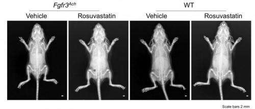
Following the success of treating TD in vitro, the investigators then examined whether similar positive effects could be made against ACH. Cultured ACH-iPS cells showed promise, so the team observed statin treatment on transgenic mice that had the FGFR3 mutation and ACH symptoms, including short bones. Sacrifice at 15 days showed that daily intraperitoneal injections of statin into these mice recovered bone length (figure 1). Furthermore, organ cultured cartilage from ACH mice treated with statin resulted in increased expression of the aforementioned three genes and also in Runx2 and Col10a1, which indicated that statin stimulated both the differentiation and maturation of ACH mouse chondrocytes. The authors also discovered that statin stimulated the degradation of FGFR3 in mice by targeting the proteasomal pathway, offering new insight on the mechanism of action.
Because statins have been long used to treat hyperlipidemia and heart disease in adults, there already exists exhaustive information about their appropriate dose and dosage. Therefore, the application of statins to TD and ACH to the clinic should be accelerated relative to normal drug development.
At the same time, Prof. Tsumaki is cautious about these findings, especially with regards to children. "Currently, statins should not be used in children, because statins decrease cholesterol, an essential steroid for the growth and development of children". He adds that further examination is also needed on the appropriate delivery and dose for skeletal dysplasia treatment. "We show that an injection of 1 mg per kg of rosuvastatin into the ACH mouse model restored bone growth in the limbs and head. However, this dose would translate into 70 mg per day for a 70 kg human, which far exceeds the 20 mg limit in Japan and 40 mg limit in Europe and the U.S".
"Because statins are used primarily in adults, there is a paucity of data for child and infant patients. Therefore, much more study on the efficacy and safety of statins, especially in the young, is needed before they reach the clinical stage for TD and ACH treatment".
CiRA Director and Professor Shinya Yamanaka, the pioneer of iPSC technology commented; "This study is an important research result, as it demonstrates that patient-specific iPSCs can be an effective tool for drug repositioning. I hope this approach will be a model to help us find new cures for other intractable disease".
More information: Statin treatment rescues FGFR3 skeletal dysplasia phenotypes, Nature, dx.doi.org/10.1038/nature13775


