March 28, 2016 report
Researchers produce stunning 4D map of somatosensory processing in the brain

(Medical Xpress)—It goes without saying that the brain is an unbelievably complex piece of biological architecture, but the depth of that complexity is often unaddressed. Applying network theory to the brain has clarified many neural functions and led to the discovery that brain structures function as hubs for a number of interrelated brain networks. But to date, no fine-grained map of the brain's spatiotemporal dynamics exists.
Consider microprocessors: Current technologies rely on increasingly small, dense microarchitectures in two dimensions. As conventional photolithography reaches the limits imposed by physics, it becomes increasingly difficult to shrink two-dimensional microarchitecture, and chip designers are pursuing new paradigms. One is three-dimensional microarchitecture, which layers stacks of two-dimensional chips and interconnects them vertically.
The brain also has such a layered 3D architecture, but it is millions of times more complicated. Because of the difficulties inherent in observing neural dynamics, it is unknown how exactly those layers interact with each other and whether cortical dynamics use parallel or serial operations.
In a startling new study reported in the Proceedings of the National Academy of Sciences, a collaborative of researchers in Italy and Germany have combined anatomical and functional data from intracerebral recordings of nearly 100 patients suffering from drug-resistant epilepsy who had been stereotactically implanted with intracerebral electrodes during surgical evaluation.

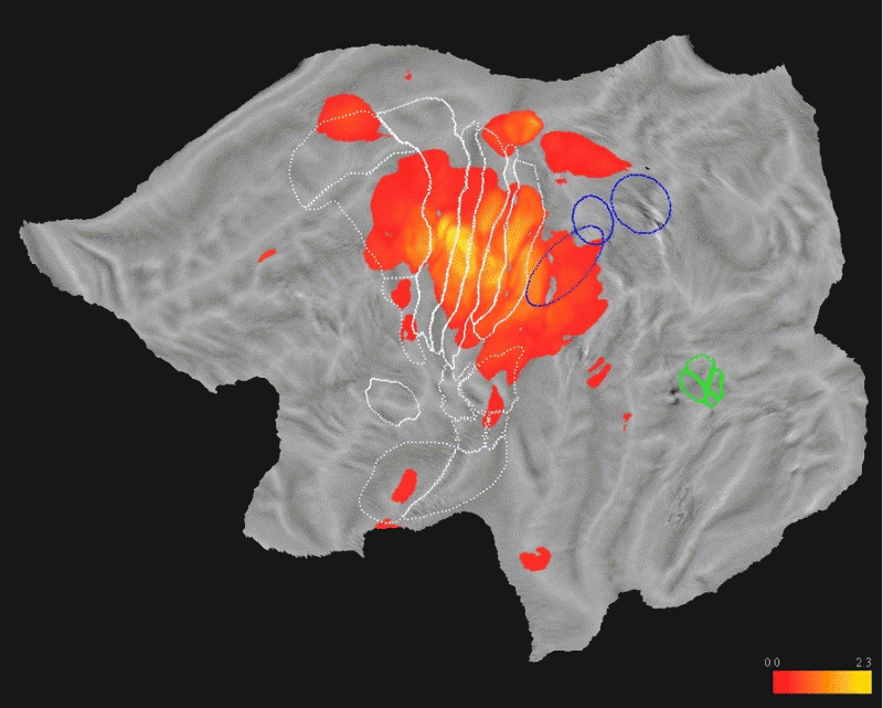
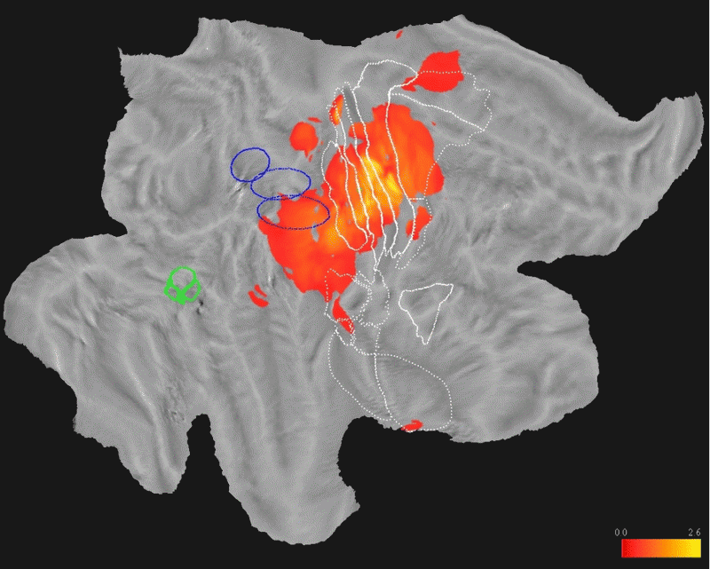
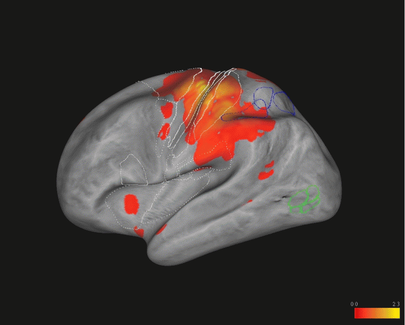
From this wealth of difficult-to-obtain data, the researchers produced four-dimensional maps of human cortical processing of nonpainful somatosensory stimuli over time and across brain regions. Astonishingly, the maps reveal that the human somatosensory system devoted to the hand alone encompasses a widespread network across more than 10 percent of the cortical surface of both brain hemispheres.
The network includes components associated with the primary somatosensory cortex, as well as motor, premotor and inferior parietal regions. But the researchers were surprised to discover that it also included optical components. They write, "...we were able to show that, indeed, somatosensory information reaches the putative ventral part of the medial superior temporal area, suggesting a possible integration between tactile and visual information... this integration may serve to control tracking of moving objects not only with the eyes... but also with arms."
The phasic, or temporal, component of the data provided evidence of the brain's cortical processing mode, and according to the researchers, it favors the parallel processing of sensory information between two of the studied regions. But they caution that the 10 millisecond frames of data that they studied are not fine-grained enough to fully confirm this conclusion and that further investigation is required.

Finally, the authors conclude that the insular cortex is consistently activated by stimulation of the hand's median nerve, and aligns with existing conclusions that this part of the insula is specifically adapted for somatosensory processing. "Our finding of a tonic activity of this sector may be related to the involvement of the posterior insula in the integration of somatosensory inputs with other modalities," they write.
More information: Four-dimensional maps of the human somatosensory system. PNAS 2016 ; published ahead of print March 14, 2016, DOI: 10.1073/pnas.1601889113
Abstract
A fine-grained description of the spatiotemporal dynamics of human brain activity is a major goal of neuroscientific research. Limitations in spatial and temporal resolution of available noninvasive recording and imaging techniques have hindered so far the acquisition of precise, comprehensive four-dimensional maps of human neural activity. The present study combines anatomical and functional data from intracerebral recordings of nearly 100 patients, to generate highly resolved four-dimensional maps of human cortical processing of nonpainful somatosensory stimuli. These maps indicate that the human somatosensory system devoted to the hand encompasses a widespread network covering more than 10% of the cortical surface of both hemispheres. This network includes phasic components, centered on primary somatosensory cortex and neighboring motor, premotor, and inferior parietal regions, and tonic components, centered on opercular and insular areas, and involving human parietal rostroventral area and ventral medial-superior-temporal area. The technique described opens new avenues for investigating the neural basis of all levels of cortical processing in humans.
© 2016 Medical Xpress


