Brain's center for perceiving 3-D motion is identified (w/ Video)

Ducking a punch or a thrown spear calls for the power of the human brain to process 3-D motion, and to perceive an object (whether it's offensive or not) moving in three dimensions is critical to survival. It also leads to a lot of fun at 3-D movies.
Neuroscientists have now pinpointed where and how the brain processes 3-D motion using specially developed computer displays and an fMRI (functional magnetic resonance imaging) machine to scan the brain.
They found, surprisingly, that 3-D motion processing occurs in an area in the brain—located just behind the left and right ears—long thought to only be responsible for processing two-dimensional motion (up, down, left and right).
This area, known simply as MT+, and its underlying neuron circuitry are so well studied that most scientists had concluded that 3-D motion must be processed elsewhere. Until now.
"Our research suggests that a large set of rich and important functions related to 3-D motion perception may have been previously overlooked in MT+," says Alexander Huk, assistant professor of neurobiology. "Given how much we already know about MT+, this research gives us strong clues about how the brain processes 3-D motion."
For the study, Huk and his colleagues had people watch 3-D visualizations while lying motionless for one or two hours in an MRI scanner fitted with a customized stereovision projection system.
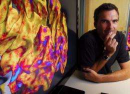
The fMRI scans revealed that the MT+ area had intense neural activity when participants perceived objects (in this case, small dots) moving toward and away from their eyes. Colorized images of participants' brains show the MT+ area awash in bright blue.
The tests also revealed how the MT+ area processes 3-D motion: it simultaneously encodes two types of cues coming from moving objects.
There is a mismatch between what the left and right eyes see. This is called binocular disparity. (When you alternate between closing your left and right eye, objects appear to jump back and forth.)
For a moving object, the brain calculates the change in this mismatch over time.
Simultaneously, an object speeding directly toward the eyes will move across the left eye's retina from right to left and the right eye's retina from left to right.
"The brain is using both of these ways to add 3-D motion up," says Huk. "It's seeing a change in position over time, and it's seeing opposite motions falling on the two retinas."
That processing comes together in the MT+ area.
"Who cares if the tiger or the spear is going from side to side?" says Lawrence Cormack, associate professor of psychology. "The most important kind of motion you can see is something coming at you, and this critical process has been elusive to us. Now we are beginning to understand where it occurs in the brain."
Huk, Cormack, and post-doctoral research and lead author Bas Rokers published their findings in Nature Neuroscience online the week of July 7.
Source: University of Texas at Austin (news : web)


