New approach simplifies Parkinson's surgery

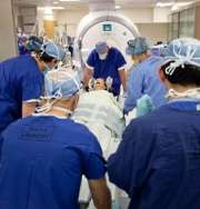
(Medical Xpress) -- University of Wisconsin Hospital and Clinics has become the second academic medical center in the country where neurosurgeons can perform deep-brain stimulation (DBS) in an intra-operative MRI (iMRI) suite.
The new operating suite allows neurosurgeons to perform faster and more accurate brain surgery while a real-time magnetic resonance image of the patient’s brain is displayed.
For patients, the result is a quicker and more comfortable experience. Conventional DBS surgery, for treatment of conditions such as Parkinson’s disease, requires that patients be conscious during procedure. In general, the new technique should cut in half the six hours needed for the older type of surgery.
"This makes DBS surgery much easier on our patients,'' says Dr. Karl Sillay, a UW Health neurosurgeon. "Previously, patients had to be awake during the surgery to help determine when the electrodes were in the correct position."
Improves the symptoms of Parkinson’s by directing electrical stimulation to a part of the brain called the subthalamic nucleus. This pea-sized region is located about six centimeters beneath the surface of the frontal cortex.
In the past, surgeons located the spot by scanning the patient before surgery. A stereotactic frame, affixed to the patient’s skull, allowed surgeons to identify the spot by use of three-dimensional coordinates. They then used micro-electrode recordings to be sure the electrode was in the correct place. Patients had to be awake and able to respond to the surgeon for this technique to work correctly. They also had to stop taking their anti-Parkinson’s medication 12 hours before surgery.
In the new iMRI suite, surgeons use the image from the iMRI to guide the electrode into place. A computer system created by Surgivision uses a plastic grid on the patient’s head, coordinated with the iMRI image, to precisely guide the neurosurgeon’s placement of the electrode.
Special surgical tools, made of titanium and plastic, allow surgery inside the powerful MRI magnet.
Patients can stay on their medication and be under general anesthesia. The new technique also has a safety component - since intracranial bleeding occurs in about 2 percent of DBS procedures, surgeons can spot the problem immediately on the real-time iMRI.
The technique will get much of its use during procedures when electrodes are placed in the brain to relieve the symptoms of Parkinson's disease, essential tremor and other neurological conditions. But the iMRI operating room is also being used for other types of brain surgery.
For example, says Dr. Robert Dempsey, chair of neurosurgery, having the MRI right in the operating room will also benefit brain tumor patients.
"This superb technology provides our surgeons with additional images of the tumor while the patient is undergoing surgery," Dempsey says. "This means we are able to operate with additional certainty that the entire tumor has been removed during a single surgery, and helps ensure we are doing our very best for patients in sparing healthy tissue."
UW Hospital is one of the first in the nation to offer real-time intra operative placement of DBS electrodes.
"This new system should be much superior for patients and represents a major step forward in our ability to treat complex neurological diseases," says Dr. Sillay.


