November 28, 2011 feature
Beyond brain scanning: Simultaneous high-resolution 3D neural imaging and photostimulation

(Medical Xpress) -- Neuroanatomy and neurophysiology are inherently three-dimensional domains. Neuronal cell body projections – axons and dendrites – can interconnect large numbers of neurons distributed over large cortical distances. Since the brain processes sensory, somatic, conceptual, and other classes of information in this 3D structural space, the need to (1) image neural structures and (2) stimulate and record neural signals are essential to understanding the relationship between brain structure and function. While 3D imaging and 3D photostimulation using scanning or parallel excitation methods have been used, they have not previously been combined into an optical system that can successfully decouple the corresponding optical planes when using a single lens – a shortcoming that has limited investigators to small neural areas. Recently, however, scientists at Université Paris Descartes have combined digital single photon holographic stimulation with remote-focusing-based epifluorescent functional imaging to overcome these limitations.
Researchers Francesca Anselmi and Cathie Ventalon in the Emiliani Wavefront-Engineering Microscopy Group led by Dr. Valentina Emiliani, along with Aurélien Bègue and David Ogden, work at the intersection of physics and biology. As such, they encountered several challenges in designing and performing their experimental demonstration.
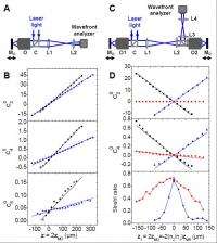
“The experimental characterization of the remote focusing system required a very precise analysis of optical aberrations,” says Emiliani. On the practical side, this meant that the setup had to be very carefully aligned. “We used a wavefront analyzer to estimate optical aberrations at different points along the light path, which helped us optimize system alignment. We were also very careful when choosing the optical elements.” For example, the team substituted conventional dichroic mirrors (used to separate excitation from fluorescent light) with thicker ones (5 mm), in order to reduce astigmatism.
A general problem when developing new technologies for neuroscience is to guarantee the necessary experimental stability while operating with a setup under development. They therefore tried to standardize all technical operations as much as possible in order to obtain highly reproducible conditions. “The alignment of the microscope was checked every day before the beginning of the experiment,” Emiliani notes. “The optimal excitation conditions were determined at the beginning of an experimental series – often by performing control experiments in another, more conventional, setup, equipped with electrophysiology to directly measure the electrical output of neurons. The filling of the neurons with fluorescent indicator was also carries out in the electrophysiology setup, immediately before the beginning of every optical experiment.” Given these practical constraints, the experimental day was generally long and intense, and often required the participations of more than one experimenter.
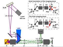
As might be expected, the team had to develop and leverage a number of insights, innovations and techniques to address these challenges. Specifically, in order to realize independent photoactivation and imaging in 3D, the team coupled two techniques: digital holography and remote focusing. Digital holography is a light patterning technique that allowed them to place multiple excitation spots simultaneously in 3D. In digital holography, the laser light used for excitation is coupled into the microscope through the principal objective, while remote focusing allows recreating an image of the biological sample in a remote location thanks to the use of a second microscope, symmetrical to the principal instrument. “The selection of the imaging plane is made at the level of this second microscope,” Emiliani explains, “and is thus completely independent of photoactivation. In particular, a blue lamp is used to uniformly illuminate the sample and excite fluorescence. A perfect fluorescent image of the sample is created by the remote microscope, and a mirror is then moved through this image in order to select the final imaging plane, as the light is reflected back to the camera.”

A key aspect of this setup is that oblique imaging can be obtained if the remote mirror is tilted instead of axially translated: In this way, the final imaging plane on the camera will also be tilted with respect to the surface of the sample – and the degree of tilt can be tuned in order to follow the morphology of the targeted structure. As a result, imaging along a tilted plane is performed in a scanless widefield process.
In order to improve the capability of optical sectioning, Emiliani sees an important innovation as being the implementation of their photoactivation and imaging system for two-photon excitation. This imaging modality is currently a standard in neuroscience research and allows improving the spatial precision of imaging and photostimulation up to submicrometer scales (i.e., comparable with the dimension of subcellular structures such as dendritic spines) and the penetration of excitation into living tissues. “Both the remote focusing imaging system and the digital holography photoactivation system have been separately implemented for two-photon excitation, so this evolution of our combined setup would allow simultaneous imaging and photoactivation with increased spatial precision deep into brain tissues.”
Another important point, she adds, would be to transform the optical prototype into a standardized setup, ready to use by biologists. “In this sense, it would be important to couple our technique with electrophysiology, in order to allow at the same time optical and electrical recording of neuronal activity.”
“In addition, we’d like to use our system to record, optically, electrical neuronal signals,” Emiliani concludes. Although this is possible thanks to the use of voltage dyes for functional imaging, the use of these dyes is sometimes technically problematic. “We believe that by allowing imaging over large regions, our scanless technique would permit to increase both the speed, due to being scanless, and the efficiency, derived from the possibility of spatially averaging the signal, of these recordings.”
The team’s findings have implications for a range of scanless neuroanatomical, neurophysiological and neuromolecular research. “The possibility of scanless imaging along a tilted plane will be important to perform fast functional imaging of neuronal activity over large areas, and several advantages can be imagined,” Emiliani agrees. “Up until now, scanless widefield imaging has been limited to structures lying on the focal plane of the microscope objective. Our system allows following the morphology of an elongated neural structure into the tissue, thus enlarging the range of structures accessible to functional imaging.” As shown in their proof of principle experiments, this flexible configuration allows recording precise information on dendritic integration – that is, the mechanisms with which neurons process and elaborate information. “Advancing our knowledge in this sense would help understanding the basic cellular units of brain functioning.”
The possibility of combining functional imaging with photostimulation in the same setup also gives them the possibility to not only monitor but also modulate neuronal activity. “Importantly, we’re able to access 3D structures. This will permit to perform causal experiments, perturbing the normal activity of neurons and observing the reaction of the system. The all-optical nature of our technique permits to reduce the invasiveness of the process, which is essential to work in physiological conditions.”
In the paper, the researchers concentrated our attention on the study of dendritic properties, given the importance of this research field for basic neuroscience. However, Emiliani points out that their setup for photoactivation and imaging could also be used to study neuronal circuits. “Photoactivation by digital holography allows us to stimulate or inhibit the activity of multiple neurons simultaneously in 3D. At the same time, the remote focusing system could be used to record functional responses from the same or other neurons in the circuit. Causal experiments can be imagined where the activity of specific neurons is perturbed and the consequences are observed in the circuit.” Again, this could be done with reduce invasiveness, thanks to the use of light for both stimulation and recording.
Another possibility is transitioning to an in silico model. “Experimental studies on dendritic integration are often combined to in silico models of neuronal functioning,” notes Emiliani. “In this sense, our system could help collecting new sets of information – in particular, relating dendritic morphological and functional properties – that could be an important input for computer models.”
Regarding application to the study of dendritic properties demonstrated in their experiments and the related study of functional connectivity in neuronal circuits, Emiliani stresses that the implementation of their setup for two-photon excitation discussed above would improve performance in both these fields through increased stimulation spatial precision, imaging resolution, and targeting larger volumes through deeper penetration of light into brain tissue.
More information: Three-dimensional imaging and photostimulation by remote-focusing and holographic light patterning, PNAS published online before print November 9, 2011, doi: 10.1073/pnas.1109111108
Copyright 2011 PhysOrg.com.
All rights reserved. This material may not be published, broadcast, rewritten or redistributed in whole or part without the express written permission of PhysOrg.com.


