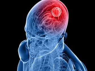
Scientists make brain tumours glow
Stereotactic needle biopsies are an established standard procedure in the diagnostic identification of brain lymphomas and certain brain tumours (gliomas). Up until now the tissue samples removed had to be examined for tumour cells in the neuropathology department whilst the operation was underway. In addition, several biopsies were often necessary to achieve diagnostic reliability and these involved several tissue extractions. With the 5-ALA fluorescence marker the correct place to make the extraction for the tumour biopsy, and thus the precise diagnosis, can be confirmed immediately in the operating theatre. This is demonstrated by a current study at the University Department of Neurosurgery of the MedUni Vienna/Vienna General Hospital. To do this scientists are getting brain tumours to glow.
The Vienna General Hospital patient has to take the 5-aminolevulinic acid (5-ALA), a fluorescent marker in powder form, dissolved in water approximately four hours before the operation. Due to the disruption of the blood-brain barrier (this is the physiological barrier between the circulatory system of the blood and the central nervous system) together with suspected enzyme defects in the tumour cell, the 5-ALA selectively concentrates there. During the surgery a blue light is emitted from the operation microscope, which causes the tumour cells to fluoresce red when 5-ALA is in use.
So far 5-ALA has already been used successfully in the neurosurgical removal of aggressive brain tumours (glioblastomas). "We have now proved in our study that it is also useful and efficient when taking a biopsy," says the author of the study, Georg Widhalm. "With clear 5-ALA fluorescence in the tumour sample the biopsy can now be finished without a neuropathological examination during the surgery and without having to carry out a serial biopsy. This results in a markedly reduced time for the operation and increased interventional safety."
It was also possible for the first time to prove that only aggressive tumour parts glow in brain tumours. This enables the removal of the tumour sample precisely in the right place, the so-called hot spot – not only when removing tumours but also when performing a biopsy on them.
As a result, it is possible to determine the classification of the degree of the tumour accurately in line with the World Health Organisation (WHO) classification. Says Stefan Wolfsberger, head of the study: "If there is a WHO grade III tumour, which is one that is growing rapidly, one can immediately put the right treatment in place after the neurosurgical intervention." This ranges from chemo to radiotherapy with the aim of maximising the life expectancy of those affected.
The study, which has now been published in the specialist journal Neurosurgical Review, is based on interdisciplinary collaboration between neurosurgery, neuropathology, oncology, radiotherapy and the radio diagnostics departments with the Comprehensive Cancer Center (CCC) at the Medical University of Vienna and the Vienna General Hospital.
More information: Widhalm, G. et al. Strong 5-aminolevulinic acid-induced fluorescence is a novel intraoperative marker for representative tissue samples in stereotactic brain tumor biopsies, Neurosurgical Review 2012; 35 (3): 381-91.


