March 10, 2014 report
The encoding of color categories in brain

(Medical Xpress) —The spectrum of visible light, like that of audible sound, spans a range of frequencies that is easy to define. There is both a minimum, and a maximum, that we can perceive. Our perception of color, while continuous, naturally partitions itself into categories that defy any sense of orderly progression, almost as if they are some kind of strange smells. Indeed if we combine the blue from the top end of the spectrum, with red from the bottom, we get a purple that proudly asserts its own uniqueness and right to be. A recent paper published in PNAS describes the use of MRI to probe the categorical representation of color in the brain. The authors conclude that the the spectral distances—how far apart different hues are from one another—are encoded in different areas then color catagories themselves.
Each ear senses sounds with a smoothly-varying bank of detector cells perhaps 3500 strong. As far as we can tell, each cell reports on just a narrow frequency segment of the contiguous whole. The nose on the other hand, has around 40 million detector cells which come in about 1000 different varieties. While it has been suggested that the nose can sense the vibratory motions of molecules, there is no known orderly representation to the 10,000 or more different types of molecules we can sense. Each scent seems to be plucked whole from an olfactory food mart, so to speak.
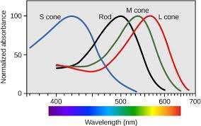
The eyes are even more extreme in their partitioning of sensory space. In round numbers, they have around 100 million rod cells for basic service, and about 10 million cones for color. By contrast with the other senses, the cones come in only about three or four different flavors which broadly overlap spectral sensitivity. The "short" cones detect the blues while the "long" cones the reds. Surprisingly, the "medium," or M cones have a maximum response that is not too far away from the L cones, and actually not all that much different from the that of the rods. How then, do we get such a crisp color experience?
The PNAS paper does not deliver us this Holy Grail, but it does suggest some ideas worth investigating a little closer. In particular, the authors report that the middle frontal gyrus, and even the cerebellum, showed more activation in a task where the color stimuli differed by catagory, rather than just different colors within catagories. This bilateral activation for catagories might be surprising considering previous studies showed a lateralized activation in the left posterior temporoparietal regions, an area associated with language or names.
While color science has been around for a while, definitions and terms are still being honed today. A new formula to calculate hue was in fact just published the other week, which corrects an oversight in the traditional formulation. Hue itself can be given tint by adding white, shade by adding black, or tone by adding grey. This same sense of incompleteness applies also to our understanding of the early-stage color processing that takes place in the strictly visual cortical areas at the back of the brain.
Semir Zeki is a well-known vision researcher who has made notable contributions to our understanding of color vision in primates. Since his original mapping studies, other researchers have made a few anatomical discoveries—in particular, the so-called color "globs," which are millimeter-sized nodules in the part of the parvocellular (small cell) stream known as the V4 complex. Today Zeki has moved on to higher things and has just published a paper on the neural correlate of the experience of mathematical beauty. That may be an appropriate topic for a researcher at the apex of their career, but it seems we still have much to learn about the more mundane experience of color.
More information: Categorical encoding of color in the brain, PNAS, www.pnas.org/cgi/doi/10.1073/pnas.1315275111
Abstract
The areas of the brain that encode color categorically have not yet been reliably identified. Here, we used functional MRI adaptation to identify neuronal populations that represent color categories irrespective of metric differences in color. Two colors were successively presented within a block of trials. The two colors were either from the same or different categories (e.g., "blue 1 and blue 2" or "blue 1 and green 1"), and the size of the hue difference was varied. Participants performed a target detection task unrelated to the difference in color. In the middle frontal gyrus of both hemispheres and to a lesser extent, the cerebellum, blood-oxygen level-dependent response was greater for colors from different categories relative to colors from the same category. Importantly, activation in these regions was not modulated by the size of the hue difference, suggesting that neurons in these regions represent color categorically, regardless of metric color difference. Representational similarity analyses, which investigated the similarity of the pattern of activity across local groups of voxels, identified other regions of the brain (including the visual cortex), which responded to metric but not categorical color differences. Therefore, categorical and metric hue differences appear to be coded in qualitatively different ways and in different brain regions. These findings have implications for the long-standing debate on the origin and nature of color categories, and also further our understanding of how color is processed by the brain.
© 2014 Medical Xpress


