May 5, 2014 report
EyeWire gamers help researchers understand retina's motion detection wiring

(Medical Xpress)—A team of researchers working at MIT has used data supplied by gamers on EyeWire to help explain how it is that the retina is able to process motion detection. In their paper published in the journal Nature, the team describes how they worked with gamers at EyeWire and then used the resulting mapped neural networks to propose a new theory to describe how it is the eye is able to understand what happens when something moves in front of it.
Scientists have known for quite some time that light enters the eye and strikes the back of the eyeball where photoreceptors respond. Those photoreceptors send information they receive to another type of neural cell known as bipolar cells—they in turn convert received signals to another signal format which is then sent to what are known as starburst amacrine cells (SACs). Signals from the SAC are sent via the optic nerve to the brain. And while scientists believe they have a pretty good idea about how the whole process works for static images, they have not been able to get a handle on what happens when images sent to the eyeball have information about things that are moving. In this new effort, the researchers sought to do just that—via assistance from thousands of gamers on the EyeWire game playing site.
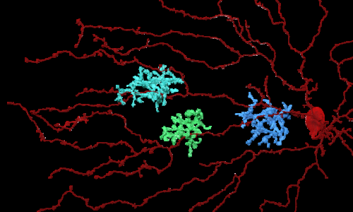
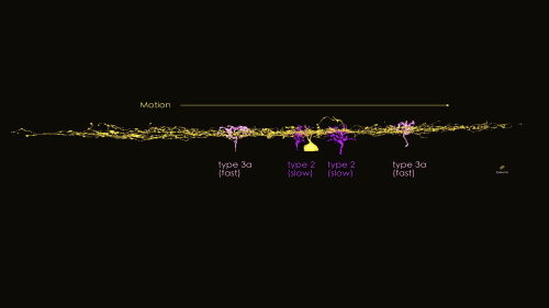
The problem with figuring out how nerve cells work in the eye, of either mice or humans, is the inability to watch what happens in action—everything is too tiny and intricate. To get around that problem, researchers have been building three dimensional models on computers. But even that gets untenable when considering the complexity and numbers of nerves involved. That's where the EyeWire gamers came in, a game was created that involved gamers creating mouse neural networks—the better they were at it the more points they got. And only the best at it were invited to play. The result was the creation of a model that the researchers believe is an accurate representation of the cells involved in processing vision, and the networks that are made up of them. From that point, on the rest was up to the research team. They noted that in the model, there were different types of bipolar cells connecting to SACs—some connected to dendrites close to the cells center, and others connected to dendrites that were farther away. Prior research had shown that some bipolar cells take longer to process information than others. The researchers believe that the bipolar cells that connect closer to the center are of the type that take longer to process signals. This, they contend, could set up a scenario where the center of the SAC receives information from both types of bipolar cells at the same time—and that, they suggest, could be how the SAC comes to understand that motion—in one direction—is occurring.
The researchers suggest their theory can be real-world tested in the lab, and expect other teams will likely do so. If they are right, the mystery of how our eyes detect motion will finally be solved.

More information: 1. Space–time wiring specificity supports direction selectivity in the retina, Nature (2014) DOI: 10.1038/nature13240
Abstract
How does the mammalian retina detect motion? This classic problem in visual neuroscience has remained unsolved for 50 years. In search of clues, here we reconstruct Off-type starburst amacrine cells (SACs) and bipolar cells (BCs) in serial electron microscopic images with help from EyeWire, an online community of 'citizen neuroscientists'. On the basis of quantitative analyses of contact area and branch depth in the retina, we find evidence that one BC type prefers to wire with a SAC dendrite near the SAC soma, whereas another BC type prefers to wire far from the soma. The near type is known to lag the far type in time of visual response. A mathematical model shows how such 'space–time wiring specificity' could endow SAC dendrites with receptive fields that are oriented in space–time and therefore respond selectively to stimuli that move in the outward direction from the soma.
2. EyeWire blog.
© 2014 Medical Xpress


