Scanning for health and disease

Many mysteries still surround the human body. In particular, the molecular and cellular processes of the body's systems and organs, and their occasional malfunctions, remain largely unobserved at the macroscale. But RIKEN researchers are shedding light on these mysteries by developing advanced imaging techniques that permit the real-time observation of intricate internal processes.
In 2013, RIKEN merged its previously isolated research initiatives in molecular imaging into one central organization dedicated to a holistic, biological understanding of health and pathology. Techniques developed by the Division of Bio-Function Dynamics Imaging at the RIKEN Center for Life Science Technologies (CLST) could revolutionize both pre-emptive medicine, which seeks to identify incipient diseases at the molecular level well before they reach the stage of manifesting recognizable symptoms, and regenerative medicine, which harnesses the body's innate healing ability to restore optimum health.
A game of tag
To achieve this goal, researchers at the division take advantage of state-of-the-art facilities at the RIKEN Kobe and Wako campuses. These facilities include magnetic resonance imaging (MRI) systems and computed tomography (CT) scanners that respectively use magnetic fields in conjunction with radio waves and x-rays to noninvasively penetrate the body and visualize slices of internal tissue. While positron emission tomography (PET) scanners trace the movement of radioactively labeled molecules introduced into the body, gamma-ray emission imaging (GREI) cameras—the first of their kind—can also join this game of tag, allowing biometals and bioactive molecules to be simultaneously visualized.
Some of the 14 laboratories at the Division of Bio-Function Dynamics Imaging construct these next-generation imaging devices, whereas others customize the associated markers so that specific biological phenomena, including diseased states, can be imaged. Researchers use the on-site baby cyclotrons, 'hot' labs and automated synthesizers to produce and process radioactive isotopes for PET imaging. These customized markers have greatly enhanced the usefulness of PET for monitoring the fate of drugs and the manifestation of disease in the body.

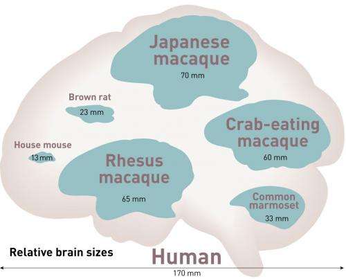
For example, to study the antidepressant effects of the anesthetic ketamine, researchers at the division radioactively labeled two molecules associated with the signaling of the neurotransmitter serotonin. They then used PET to monitor the movement of these tagged markers in the brains of conscious monkeys following ketamine treatment. From the resulting scans, they identified increased activity in two brain regions involved in regulating the reward response. Similarly, other researchers at the division tagged molecules associated with neuroinflammation in patients suffering from chronic fatigue syndrome, which helped identify a link between inflammation and the debilitating disease.
Pre-empting disease
The division also supports several major national-level initiatives, including a molecular imaging research program for innovation in drug discovery and diagnostic technology, a project to advance cell replacement therapies for Parkinson's disease using induced pluripotent stem cells, and a large-scale, neuroimaging-based study of Alzheimer's disease.
Technologies developed at the division will expand and enhance the clinician's toolkit by providing new drugs and an extensive palette of specialized biomarkers for identifying the onset of various diseases prior to the appearance of symptoms. Ultimately, bioimaging tools could demystify the human body in the same way that satellites have done for the Earth.


