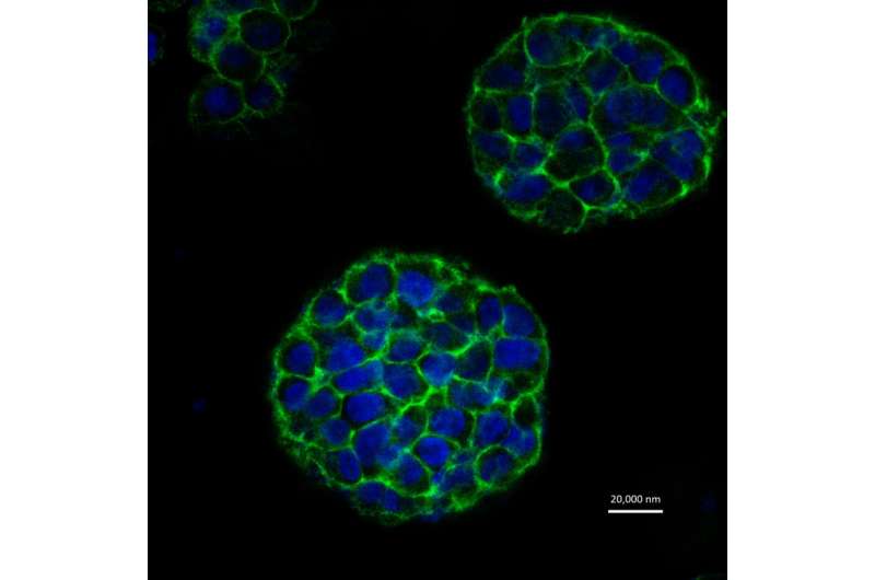
3D printer generates realistic model of a cancerous tumour

An international scientific team has successfully created a three-dimensional model of a cancerous tumour using a 3D printer. Their model could ultimately help discover new drugs and cast new light on how tumours develop, grow and spread.
Presented in the IOP Publishing journal Biofabrication, the realistic 3D model consists of a scaffold of fibrous proteins coated in cervical cancer cells. A 10mm by 10mm grid structure – made from gelatin, alginate and fibrin – recreates the fibrous proteins that make up the extracellular matrix of a tumour. The grid structure is coated in Hela cells: a unique, "immortal" cell line that was originally derived from a cervical cancer patient in 1951.
The most effective way of studying tumours is in a clinical trial. However, ethical and safety limitations make it difficult for such studies to be carried out on a wide scale. To overcome this, two-dimensional models – consisting of a single layer of cells – have been created to mimic the physiological environment of tumours and test anti-cancer drugs in a realistic way.
With the advent of 3D printing, it is now possible to provide a more realistic representation of the environment surrounding a tumour. The researchers demonstrated this by comparing results from their 3D model with results from a 2D model.
After testing if the cells remained viable, or alive, after printing, the researchers examined how the cells proliferated, how they expressed a specific set of proteins that help tumours spread, and how resistant the cells were to anti-cancer drugs.
They found that 90% of the cancer cells remained viable after the printing process. In addition, the 3D model shared more similarities with a tumour than 2D models including a higher proliferation rate, higher protein expression and higher resistance to anti-cancer drugs.
"We have provided a scalable and versatile 3D cancer model that shows a greater resemblance to natural cancer than 2D cultured cancer cells," says the lead author, Professor Wei Sun of Tsinghua University in China and Drexel University in the United States.
The researchers are now trying to understand both cell-cell and cell-substrate communication and immune responses for their printed tumour-like models. "With further understanding of these 3D models, we plan to use them to study the development, invasion, metastasis and treatment of cancer using specific cancer cells from patients," says Professor Sun. "We can also use these models to test the efficacy and safety of new cancer treatment therapies and cancer drugs."


