Facility churns out radioactive agents to investigate the subcellular activity that drives disease

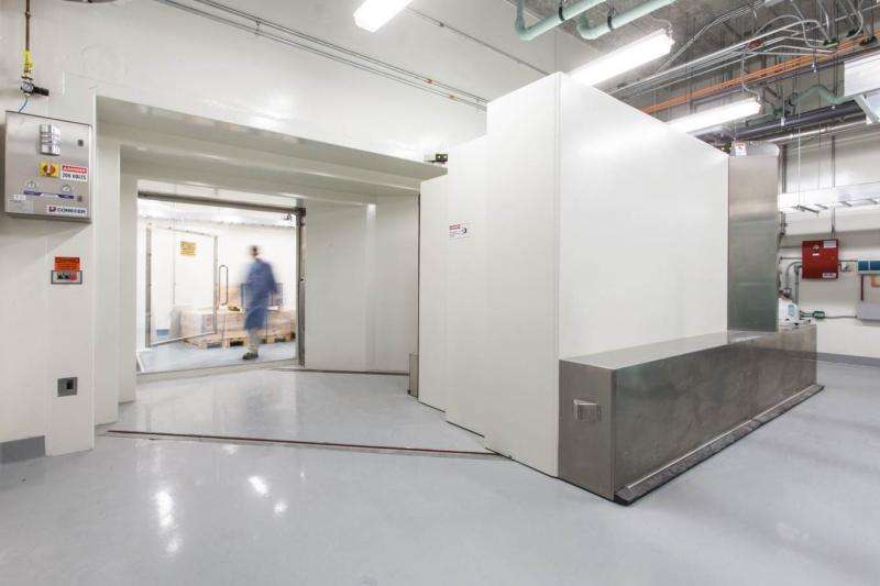
A modern-day version of the machine that smashed atoms for the Manhattan project now resides on a quiet corner of UNC's medical research campus, in a room below the basement of Marsico Hall. While its predecessor was used to conduct the physics experiments that led to the atomic bomb, this shiny new instrument is currently tasked with making radioactive tracers that could transform the fight against cancer and other complex diseases.
The device is called a cyclotron, a type of particle accelerator that laces common elements like carbon, oxygen, and nitrogen with radioactivity. UNC scientists are infusing radiation-rich products into live animal models to create better images of biological processes and diseases and to gauge the effects of new drugs, all with unprecedented molecular precision. Because this cyclotron is the only instrument on a Triangle-based academic campus that is approved to produce radiopharmaceuticals for clinical use, laboratories at Duke and NC State are also clamoring for a piece of the action.
"Most of the time when you think of imaging it brings to mind physical structures, like a section of your brain or a spot in your lung," said Hong Yuan, Ph.D., director of UNC's Small Animal Imaging Facility. "But molecular imaging goes beyond anatomy to give us a glimpse of what's happening at the subcellular level. We can tag sugar molecules and watch tumors as they use up energy. We can label dopamine receptors to probe the mechanisms of neurological diseases."
The installation of the cyclotron and the associated radiochemistry laboratory is a big enhancement to the university's molecular imaging capabilities. It will provide much more in-depth, mechanistic information to enrich research studies. Yuan's facility has been in operation since 2005 as part of the Biomedical Research Imaging Center (BRIC), which now fills three floors of Marsico Hall with instruments for magnetic resonance imaging (MRI), positron emission tomography (PET), computed tomography (CT), single positron emission computed tomography (SPECT), optical imaging, and ultrasound imaging. Each machine in the Small Animal Imaging Facility is nearly identical to those used to diagnose and treat human patients in UNC Clinics, only right-sized for small animals.

"For our imaging studies, we treat animals as if they were patients," said Yuan. "We use anesthesia to put them to sleep, we monitor their vital signs, and we scan them and process the images the same way we would if they were people."
The facility uses radiotracers to conduct molecular imaging in small animal models. However, these compounds are by design unstable and short-lived, decaying within hours or sometimes even minutes. If researchers had to order all of them by mail, some of these reagents (like Oxygen-15) would have lost their usefulness before they even arrived at the laboratory. Other radioisotopes (like Copper-64) last longer and could be ordered from other cyclotrons or commercial vendors, but even then they are expensive. Therefore, having a dedicated cyclotron on site greatly expands the types of studies researchers can explore and reduces the costs associated with research.
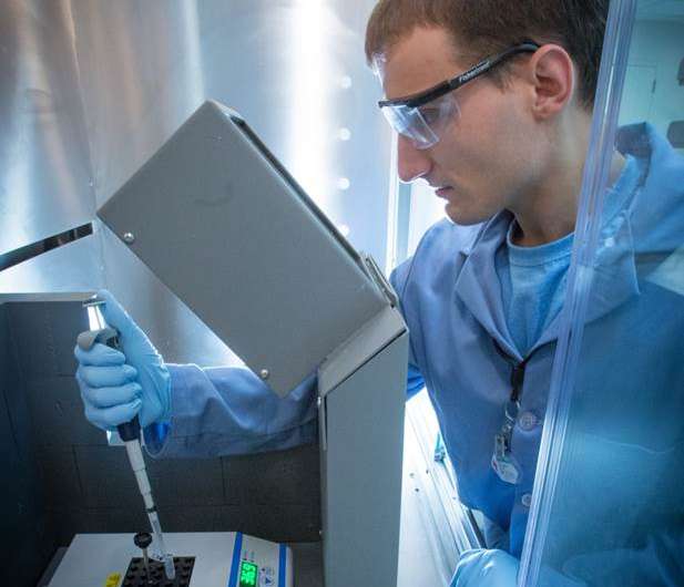
The cyclotron works by taking protons and accelerating them until they are moving at nearly a quarter of the speed of light. It then shoots the protons at the element it wants to make radioactive, like a cue ball hitting and transferring energy to its target on a pool table. The radiochemistry lab incorporates these radioactive elements or radioisotopes into chemical compounds that can then be injected into live subjects for molecular imaging. For a time, these unstable compounds continue to emit energy, which can be picked up by instruments like PET scanners. The facility plans to produce a suite of radioactive tracers for both animal and clinical imaging studies focused on cancer, neurological conditions, and cardiovascular diseases.
"One of our long term goals is to translate lab discoveries into the clinical setting, which will greatly benefit patients," said Zibo Li, PhD, director of the Cyclotron and Radiochemistry Research Program.
Because the cyclotron facility deals with a huge amount of radioactive material, special precautions are put in place to protect researchers and the public from potentially dangerous radiation exposure. The cyclotron lies between two giant slabs of concrete – each several feet thick – and its door is made of 11 tons of lead alloy. Access to the cyclotron and radiochemistry lab is only given to well-trained workers. These workers are armed with dosimeters and Geiger counters that monitor how much radiation they are exposed to at any given time. Highly sensitive foot and hand monitors guarantee no radioactivity is carried out of the facility accidentally. The facility is also equipped with a special compressed air system that captures radiative gases created during production and stores them in the cyclotron vault for decay.
The imaging center is certainly in good hands. Li has years of experience developing novel molecular imaging probes. Eric Smith, PhD, director of Radiopharmacy and Quality Management, is in charge of the regulatory issues that surround the use of radiotracers. Terri Wong, MD, director of Molecular Imaging at BRIC, is a clinician radiologist skilled at nuclear medicine. Together with Yuan, the facility staff can provide valuable expertise and support to scientists who want to use imaging tools to facilitate their own research projects.
"Basic biological researchers know that imaging can be valuable, but they often don't know how to go about it," said Yuan. "One of my responsibilities is to bridge the gap between researchers and the resources that could help them answer basic biological questions. Our imaging methods can give them information that they would never be able to gain using traditional methods like cell assays or protein assays. It has the potential to fundamentally change the way we approach research."


