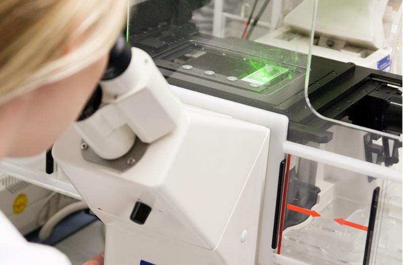
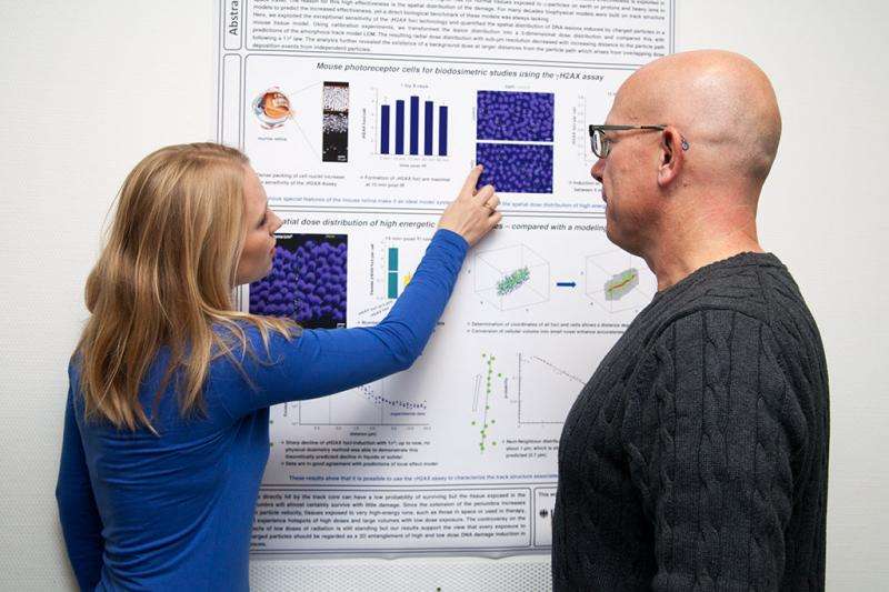
Researchers conduct high-res analyses on lesions in tissue

A precise understanding of how ion beams affect biological tissue is of great importance for both radiotherapy applications and the assessment of radioprotection risks, e.g. to astronauts on long term missions in space.
The radiation biology and biophysics research groups headed by Professor Markus Löbrich (TU Darmstadt) and Professor Marco Durante (GSI) respectively were the first to conduct experimental high resolution analyses on the 3D lesion distribution induced by high energy ion beams in biological tissue and to compare these with theoretical model predictions.
The biological effects of radiation consist in the damage caused to genetic information (DNA) contained in every cell nucleus. However, cells feature powerful repair mechanisms that can undo a lot of the damage caused by radiation. That ion beams can induce greater effects than conventional photon (e.g. X ray) radiation can be explained by the extremely high energy they emit over a very small space around the ions' path. In other words, ion beams can induce highly complex local damage that is far more resistant to repair efforts than the damage caused by photon radiation.
The conceptions favoured to date of ion beam induced 3D lesion patterns are based above all on theoretical considerations deduced from measurements of physical properties. There are no measurement data available for biological systems.

In a joint research project, scientists at the TU Darmstadt and GSI Hemholtzzentrum für Schwerionenforschung were the first to analyse 3D lesion distribution in biological tissue on the submicrometre level and to compare their findings with theoretical predictions. The radiation experiments at GSI used high energy ion beams with the same characteristics as the cosmic radiation in space.
Identification with marker
The analyses were conducted on a tissue with a particularly high density of cell nuclei, facilitating a virtually continuous detection of DNA damage. The identification of damage involved the use of a marker for the most serious form of biological damage, the DNA double strand break, causing the irreversible loss of key genetic information.
This experimental approach can visualise the traces of ion induced DNA damage over many cells. The measurements show clearly the concentration of damage at the centre of the ion path and a rapidly declining lesion frequency away from this.
Effects predicted to greater precision
On the one hand, these biological findings confirm the assumptions of 3D lesion distribution based on measured physical properties. On the other, they can be used for a critical analysis and quasi calibration of the various prediction models. These data provide an essential constituent of a model for the prediction of radiation efficacy that was developed by GSI physicists and applied for treatment planning at the ion beam therapy centres in Heidelberg, Marburg, Pavia, and Shanghai for their tumour treatment schedules.
More information: "Direct measurement of the 3-dimensional DNA lesion distribution induced by energetic charged particles in a mouse model tissue." PNAS 2015 ; published ahead of print September 21, 2015, DOI: 10.1073/pnas.1508702112


