New microscopic technique could help detect, diagnose metastatic melanomas
The fight against skin cancer just got a new weapon. For years, melanoma researchers have studied samples that were considered uniform in size and color, making them easier to examine by more conventional means. But melanomas don't always come in the same shape and hue; often, melanomas are irregular and dark, making them difficult to investigate. Now, researchers at the University of Missouri have devised a new tool to detect and analyze single melanoma cells that are more representative of the skin cancers developed by most patients. The study, recently reported in Analyst published by the Royal Society of Chemistry, outlines the new techniques that could lead to better and faster diagnoses for the life-threatening disease.
"Researchers often seek out the types of cancerous cells that are homogenous in nature and are easier to observe with traditional microscopic devices," said Luis Polo-Parada, an Associate Professor of Medical Pharmacology and Physiology and an investigator at Mizzou's Dalton Cardiovascular Research Center. "Yet, because the vast amount of research is conducted on one type of cell, it often can lead to misdiagnosis in a clinical setting."
The team that included Gary Baker, an assistant professor of chemistry in the MU College of Arts and Science and Gerardo Gutierrez-Juarez, a professor and investigator at the University of Guanajuato in Mexico, decided to supplement an emerging technique called photoacoustic (PA) spectroscopy, a specialized optical technique that is used to probe tissues and cells non-invasively. Current systems use the formation of sound waves followed by the absorption of light which means that the tissues must adequately absorb the laser light. This is why, up until now, researchers have focused only on strong-light-absorb cells melanoma cells, Polo-Parada said.
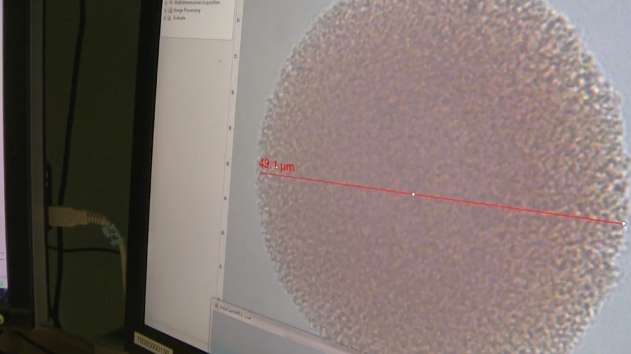
The team modified a microscope that was able to merge light sources at a range conducive to observing the details of single melanoma cells. Using the modified system, human melanoma and breast cancers as well as mouse melanoma cells were diagnosed with greater ease and efficiency. The team also noted that as the cancer cells divided, they grew paler in color but the system was able to detect the newer, smaller cells as well.
"Overall, our studies show that by using modified techniques we will be able to observe non-uniform cancer cells, regardless of their origin," Polo-Parada said. "Additionally, as these melanoma cells divide and distribute themselves throughout the blood, they can cause melanomas to metastasize. We were able to observe those cancers as well. This method could help medical doctors and pathologists to detect cancers as they spread, becoming one of the tools in the fight against this fatal disease."
The study is titled "Spectrophotometric analysis at the single-cell level: elucidating dispersity within melanic immortalized cell populations."
More information: Luis Polo-Parada et al. Spectrophotometric analysis at the single-cell level: elucidating dispersity within melanic immortalized cell populations, The Analyst (2017). DOI: 10.1039/C6AN02662A


