Newborns with CHD show signs of brain impairment even before cardiac surgery

Survival rates have soared for infants born with congenital heart disease (CHD), the most common birth defect, thanks to innovative cardiac surgery that sometimes occurs within hours of birth. However, the neurodevelopmental picture for these infants has remained stubbornly unchanged with more than 50 percent experiencing neurodevelopmental disabilities.
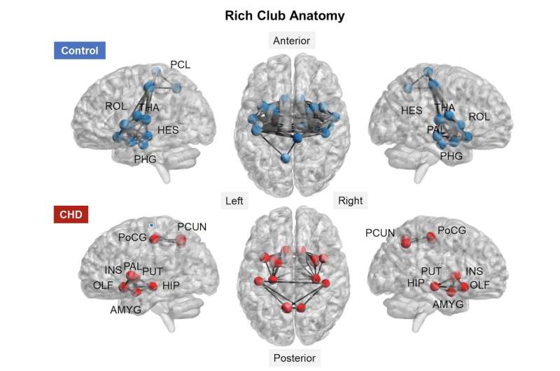
Using a novel imaging technique, Children's National Health System researchers demonstrate for the first time that the brains of these high-risk infants already show signs of functional impairment even before they undergo corrective open heart surgery. Looking at the newborns' entire brain topography, the team found intact global organization—efficient and effective small world networks—yet reduced functional connectivity between key brain regions.
"A robust neural network is critical for neurons to travel to their intended destinations and for the body to carry out nerve cells' instructions. In this study, we found the density of connections among rich club nodes was diminished, and there was reduced connectivity between critical brain hubs," says Catherine Limperopoulos, Ph.D., director of the Developing Brain Research Laboratory at Children's National and senior author of the study published online Sept. 28, 2017 in NeuroImage: Clinical. "CHD disrupts how oxygenated blood flows throughout the body, including to the brain. Despite disturbed hemodynamics, infants with CHD still are able to efficiently transfer neural information among neighboring areas of the brain and across distant regions."
The research team led by Josepheen De Asis-Cruz, M.D., Ph.D., compared whole brain functional connectivity in 82 healthy, full-term newborns and 30 newborns with CHD prior to corrective heart surgery. Conventional imaging had detected no brain injuries in either group. The team used resting state functional connectivity magnetic resonance imaging (rs-fcMRI), a imaging technique that characterizes fluctuating blood oxygen level dependent signals from different regions of the brain, to map the effect of CHD on newborns' developing brains.
The newborns with CHD had lower birth weights and lower APGAR scores (a gauge of how well brand-new babies fare outside the womb) at one and five minutes after birth. Before the scan, the infants were fed, wrapped snugly in warm blankets, securely positioned using vacuum pillows, and their ears were protected with ear plugs and ear muffs.
While the infants with CHD had intact global network topology, a close examination of specific brain regions revealed functional disturbances in a subnetwork of nodes in newborns with cardiac disease. The subcortical regions were involved in most of those affected connections. The team also found weaker functional connectivity between right and left thalamus (the region that processes and transmits sensory information) and between the right thalamus and the left supplementary motor area (the section of the cerebral cortex that helps to control movement). The regions with reduced functional connectivity depicted by rs-fcMRI match up with regional brain anomalies described in imaging studies powered by conventional MRI and diffusion tensor imaging.
"Global network organization is preserved, despite CHD, and small world brain networks in newborns show a remarkable ability to withstand brain injury early in life," Limperopoulos adds. "These intact, efficient small world networks bode well for targeting early therapy and rehabilitative interventions to lower the newborns' risk of developing long-term neurological deficits that can contribute to problems with executive function, motor function, learning and social behavior."
More information: Josepheen De Asis-Cruz et al, Aberrant brain functional connectivity in newborns with congenital heart disease before cardiac surgery, NeuroImage: Clinical (2017). DOI: 10.1016/j.nicl.2017.09.020


