Ear's inner secrets revealed with new technology
 and the auditory bones the malleus (light yellow), the incus (orange) and the stapes (blue). The green spiral at the bottom is the cochlea’s auditory membrane. There are hair cells on it that catch vibrations from the ear drum and the auditory bones. The yellow structure is the auditory nerve. Credit: Springer Nature")
What does it actually look like deep inside our ears? This has been very difficult to study as the inner ear is protected by the hardest bone in the body. But with the help of synchrotron X-rays, it is now possible to depict details inside the ear three-dimensionally. Together with Canadian colleagues, researchers from Uppsala University have used the method to map the blood vessels of the inner ear.
The study, which was published in the scientific journal Scientific Reports, can provide an explanation for why it is so effective to treat deafness in people with cochlear implants (CI). This is a method that means that an electrode that electrically stimulates the auditory nerve is operated into the inner ear. To-date, around 500,000 people worldwide have been treated with this technique. In Uppsala, the operation is also performed on patients with severe hearing loss, but who can perceive sounds with lower frequencies.
"We need to get better at understanding the micro-anatomy of the human auditory organ and how electrodes operated in affect structures in the cochlea. It can lead to an improved electrode design and better hearing results. 3-D reconstructions mean that we can study new surgical paths to the auditory nerve," says Helge Rask-Andersen, Senior Professor in Experimental Otology at the Department of Surgical Sciences.
To be able to study the blood vessels in the inner auditory organ, the researchers used the synchrotron system in Saskatoon, Saskatchewan, Canada. The system, which is one of eight in the world, is as large as a football pitch and accelerates particles with very high energy. This makes it possible to create pictures of the smallest parts of the inner ear. Through computer processing, the images can then be made three-dimensional.
-
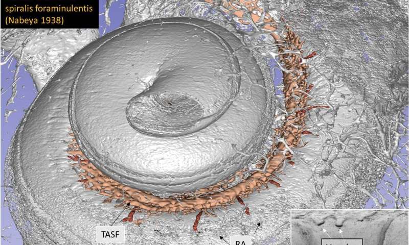
The cochlea and its blood vessel canals. In the cochlea, there are 15,000 hair cells that catch the vibrations (sound waves) from the ear drum and the auditory bones. Credit: Springer Nature -
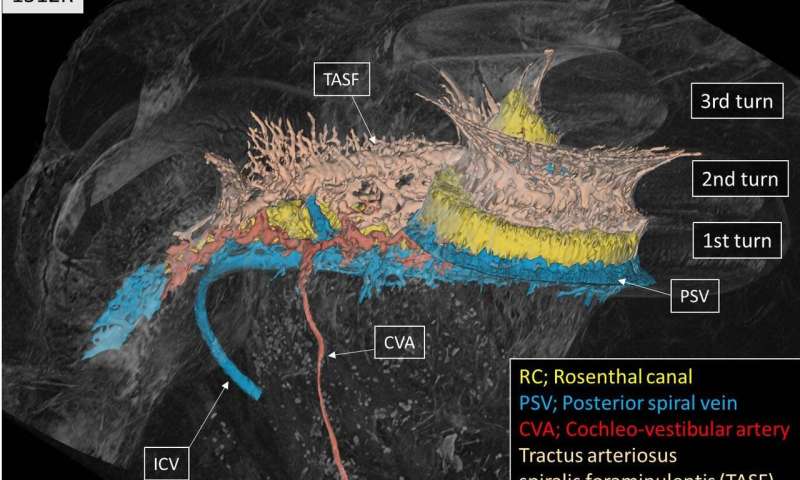
The small blood vessel that enters the cochlea (called the cochleo-vestibular artery, CVA). Credit: Springer Nature
The researchers hope the method in the future can contribute to new knowledge about diseases of the ear, such as Meniere's disease, sudden deafness and tinnitus, the causes of which are still largely unknown. But as yet, it is not possible to study living patients with this technique. The radiation is too strong.
"We study specimens from the deceased, meaning donated temporal bones. We hope that the technology can be modified in the future to achieve better resolution than today," says Helge Rask-Andersen.
More information: Xueshuang Mei et al. Vascular Supply of the Human Spiral Ganglion: Novel Three-Dimensional Analysis Using Synchrotron Phase-Contrast Imaging and Histology, Scientific Reports (2020). DOI: 10.1038/s41598-020-62653-0


