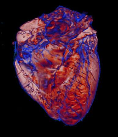
Team uses imaging to study ways the heart is affected by coronavirus

Vanderbilt University Medical Center investigators are using imaging and diagnostic pathology to examine postmortem hearts donated by victims of COVID-19. They are looking for blood clots, vascular damage and inflammation to gain a better understanding of how the coronavirus that causes COVID-19 affects the heart.
"We were hearing anecdotal stories and reports from other centers that individuals with COVID-19 suffer from hypercoagulability, microthrombi (clots in the small blood vessels), and inflammation of the blood vessel wall," said Meghan Kapp, MD, assistant professor of Pathology, Microbiology and Immunology. "I can examine blood vessels under the microscope, but getting a tissue biopsy is invasive, so I tried to think of another way to look for microinfarcts (caused by clots) or bleeds that might explain the clinical presentation of these patients."
Kapp was familiar with the research of Allison Griffin, a graduate student working with Manus Donahue, Ph.D., professor of Radiology and Radiological Sciences. Griffin and Donahue are conducting postmortem imaging of donated brains to study neurodegenerative and cerebrovascular disorders.
Griffin had previously developed radiology-pathology co-localization methods at the National Institute of Neurological Disorders and Stroke to investigate microbleeds and microinfarcts in brain tissue.
A conversation between Kapp and Griffin turned into a collaborative team effort to image hearts with MRI and CT and study the histopathology of heart tissue sections.
"We anticipate that postmortem imaging will demonstrate small vessel pathology and myocardial inflammation that correlates with pathological and clinical findings, which will help us understand how this virus is affecting the heart," Kapp said.
"I'm excited to use imaging and pathology co-localization methods to investigate a potential biomarker of COVID-19," Griffin said.
The team might make a finding on imaging that aids in improving treatments for patients with COVID-19, Kapp added.
Kapp also hopes the collaboration will extend beyond COVID-19 to other clinical scenarios such as heart transplantation or the pathologies of other organs.
"For me, this project highlights what is so tremendous about working at Vanderbilt," Kapp said. "One idea shared with a colleague led to a dynamic conversation filled with what-ifs, and we were able to rapidly bring together an amazing team of inquisitive minds and expertise from different specialties to answer our questions."


