Ultrasound-guided percutaneous needle biopsy excellent for small pleural lesions diagnosis

According to an open-access article in ARRS' American Journal of Roentgenology (AJR), ultrasound (US)-guided percutaneous pleural needle biopsy (PCPNB) has excellent diagnostic accuracy for small pleural lesions.
"US-guided PCPNB is highly likely to be diagnostic for small pleural lesions with nodular morphology on CT or US, or with pleural thickness—4.5 mm," explained Jongmin Park and colleagues from South Korea's Kyungpook National University.
To determine the diagnostic yield of US-guided PCPNB for small (? 2 cm) pleural lesions and the impact of CT and US morphologic and technical factors, Park's team retrospectively studied 103 patients (73 men, 30 women; mean age, 60.8) who underwent US-guided PCPNB of a small pleural lesion by a single experienced operator from July 2013 to December 2019. Histopathological results established final diagnosis, including from repeat US-guided and CT-guided biopsies, as well as imaging and clinical follow-up. CT and US assessed pleural morphology and thickness, while US measured needle pathway length throughout the pleura.
Summarizing their results, Park et al. noted: "[US-guided PCPNB] of small pleural lesions had a diagnostic accuracy of 85.4%. The yield was 96.4% for nodular CT lesions, 95.0% for diffuse CT lesions with thickness ?4.5 mm, 55.6% for diffuse CT lesions with thickness."
Repeating their assessments on both CT and US 2 weeks later yielded the following representative measurements:
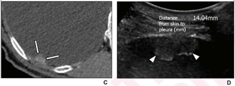
- 82-Year-Old Man Diagnosed With Tuberculous Pleurisy on US-Guided PCPNB (see image)
- 84-Year-Old Man Diagnosed with Pleural Metastasis From Lung Adenocarcinoma on US-Guided PCPNB (see image)
"In multivariable analysis," the authors of this AJR article concluded, "pleural morphology on US and needle pathway length through the pleura independently predicted diagnostic yield," adding that if the pleura is nodular or thicker than 4.5 mm on CT, US-guided PCPNB is justifiable as an initial or repeat diagnostic test.

More information: Jongmin Park et al, Ultrasound-guided Percutaneous Needle Biopsy for Small Pleural Lesions: Diagnostic Yield and Impact of CT and Ultrasound Characteristics, American Journal of Roentgenology (2020). DOI: 10.2214/AJR.20.24120


