September 20, 2021 feature
Study identifies reactive neuroinflammatory astrocyte subtypes in the mouse brain

Astrocytes are a specialized class of glial cells highly abundant in the central nervous system (CNS) of humans and other animal species. Past neuroscience research has shown that after infections, acute injuries and chronic neurodegenerative diseases, these cells can undergo an inflammatory transition. While neuroscientists have gathered substantial evidence of this transition, the factors affecting its unfolding and its many possible sub-states in different areas of the brain are still poorly understood.
Researchers at NYU Grossman School of Medicine recently carried out a study investigating the inflammatory transition of astrocytes reported in previous studies. More specifically, they investigated changes in astrocytes in the cortical region of the mouse brain after an acute inflammatory stimulus. Their findings, presented in a paper published in Nature Neuroscience, could enhance the current understanding of astrocytes and how they transform after inflammations or serious injuries.
"Recently, our lab had reported that astrocytes in the inflamed brain can be turned reactive by microglia-derived cytokines," Shane Liddelow, assistant professor at NYU Grossman School of Medicine, told Medical Xpress. "These reactive astrocytes not only become highly neurotoxic but also express a distinct gene expression profile. What we didn't know was whether these reactive genes were co-expressed in a single subtype of astrocyte, or whether we are dealing with many different astrocyte sub-states that cope differently with this inflammatory insult."
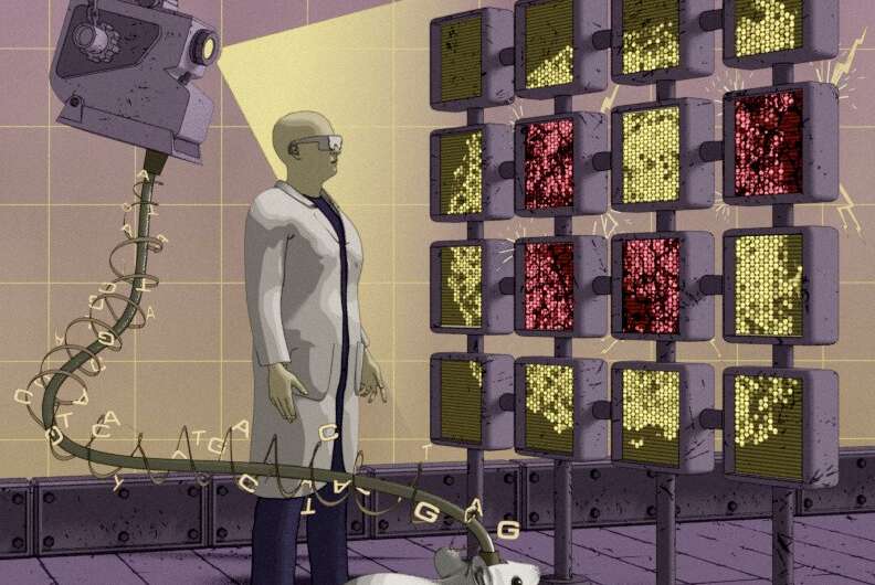
To gain a better understanding of differences in astrocytes present in the healthy and inflamed brain, Dr. Philip Hasel, lead author of the study, and his colleagues used a combination of two techniques widely employed in neuroscience research: single cell RNA-sequencing (scRNAseq) and spatial transcriptomics . Using these techniques, they were able to identify different subsets of astrocytes and understand how these subtypes respond to neuroinflammation and how this relates with their location in the brain.
"As the name implies, scRNAseq allowed us to profile the gene expression profile of single astrocytes," Hasel explained. "scRNAseq is a powerful tool to understand the cellular make up of a brain region. If you then add spatial transcriptomics, which gives you brain-region resolved transcriptome-wide gene expression, you can approximate where your healthy and inflamed astrocyte sub-states sit in the brain."
To investigate the 'inflammatory trajectory' of inflamed astrocytes, Hasel and his colleagues also created time-resolved RNA-seq data of astrocytes in mice brains that were exposed to a severe inflammatory stimulus. Their findings were highly insightful, as they revealed that a single inflammatory insult (i.e., an inflammatory event that causes damage to tissue or organs), led to several complex astrocyte sub-states with unique and dynamic gene expression profiles.
"We also found that one driver of this diverse response is the anatomical position of astrocyte subtypes," Hasel said. "More specifically, the astrocytes that responded the strongest turned out to live in strategic locations in the brain, namely near the ventricles, blood vessels and the surface of the brain."
The sequencing data collected this team of researchers is available on two easily accessible websites gliaseq.com and gliaseqPRO.com. In the future, other teams of neuroscientists could thus use this data to investigate inflammatory transitions of astrocytes further. Meanwhile, Hasel and his colleagues plan to conduct further studies aimed at better understanding the role of the reactive, 'super-responder' astrocyte subtypes they uncovered.
"Given that we know their anatomical locations, we wonder whether these astrocyte subtypes are keeping this peripheral inflammatory insult at bay or whether they're actually actively recruiting leukocytes to the locus of the insult," Hasel added. "We now have genetic tools that allow us to either track or remove certain reactive subtypes in the lab. We're excited to discover what the role of these astrocytes is in neuroinflammation."
More information: Neuroinflammatory astrocyte subtypes in the mouse brain. Nature Neuroscience(2021). DOI: 10.1038/s41593-021-00905-6
© 2021 Science X Network


