Zebrafish and AI replace some mouse experiments in cancer research

Researchers at Uppsala University have used AI to develop a new method to study brain cancer. The method is based on transplanting tumor cells from patients to fish embryos, followed by observation with AI. The method, which is described in the scientific journal Neuro-Oncology, can partly replace current mouse models for studying tumor growth and treatment.
One of the main challenges in cancer research is to study tumor growth and treatment in a relevant manner. To do this, cancer researchers often rely on experiments in mammals. A common type of experiment is based on transplanting tumor cells into mice, to characterize differences in tumor growth and therapy responses. Since mouse experiments are difficult and time consuming, and require mammals, it is important to find alternative ways.
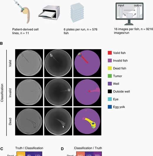
"We have developed a model in zebrafish embryos as an alternative to mouse experiments. Cells are transplanted into zebrafish and monitored for four days, and at the same time they are filmed using an automatic microscope. The film is then analyzed using AI. This is a powerful tool to measure tumor growth and to understand the effect of different drugs," says Emil Rosén, Ph.D. student at the Department of Immunology, Genetics and Pathology and one of the main authors of the study.
The researchers have used cancer cells from eleven patients with the brain tumor glioblastoma to evaluate the method. They found that cells from different patients had different survival, growth patterns and drug responses after being transplanted to the fish embryos.
"Not all cancer cells can grow in mouse models. We saw that the cells that didn't grow in zebrafish didn't grow in mice either. If the method can be used to predict how cells will function in mice, we can reduce the number of mice that are used in other studies. We have also successfully shown that the method can be employed to understand cancer metastasis and to test drugs, which makes it a promising alternative for some mouse experiments," says Professor Sven Nelander, who has led the study.
More information: Elin Almstedt et al, Real-time evaluation of glioblastoma growth in patient-specific zebrafish xenografts, Neuro-Oncology (2021). DOI: 10.1093/neuonc/noab264


