Pencil-beam scanning catheter for intracoronary optical coherence tomography

A new publication from Opto-Electronic Advances reviews a pencil-beam scanning catheter for intracoronary optical coherence tomography.
Cardiovascular disease is one of the diseases that seriously threaten human health nowadays. Its morbidity and mortality have surpassed tumors. Percutaneous coronary intervention (PCI) surgery is a kind of treatment to improve myocardial blood perfusion by using catheter technologies to open narrow or closed occlusions. In PCI surgery, optical coherence tomography (OCT) becomes more and more popular for preoperative strategy formulation and postoperative effect evaluation because of OCT's high resolution, real-time imaging speed, and label-free features.
With the promotion and application of cardiovascular OCT systems in all levels of hospitals, high performance and low-cost imaging catheters are highly demanded. The imaging catheter is the key part of intravascular OCT system and the catheter is disposable in practice. The performance of imaging catheter determines the success or failure of PCI surgery, and the imaging catheter's production cost affects the application range of cardiovascular OCT technology.
Technically, the imaging catheter consists of meters of single-mode fiber (SMF) to guide near infrared light at 1310-nm band in general and a distal focusing optics to focus the light beam onto lesions. In current mainstream gradient-index (GRIN) lens probe, a glass rod spacer is set before a GRIN lens to expand light beam for desired beam profile. This design provides great flexibility to control beam profile. However, it has several shortcomings. Firstly, the length of the glass rod spacer with several hundred micrometers is crucial and should be well controlled which is difficult to manipulate in practice. Secondly, since the light beam is expanded before GRIN lens, it is hard to fabricate small size catheters by a small GRIN lens. A large size GRIN lens is hard to be fusion spliced directly with conventional SMF and UV curing might be applied. Finally, the complicated structure increases the fabrication cost and thus set an obstacle for mass production. Consequently, how to design and fabricate a type of imaging catheter with high performance, simple architecture, and low cost is an interesting topic.
To fulfill these requirements, the research group of Associate Professor Yunxu Sun from Harbin Institute of Technology (Shenzhen) cooperated with the research group of Professor Kenneth K. Y. Wong from The University of Hong Kong and Shenzhen Vivolight Medical Device & Technology Co., Ltd. to develop a type of novel all-fiber imaging catheter for intravascular OCT application.
In this novel design, the GRIN lens was fusion spliced directly with ~1.6-m SMF, and 185-μm long no-core fiber (NCF) was fusion spliced with the GRIN fiber. NCF provides a polishing basement to make a 40-degree angle reflection surface at the tip of the probe for side-viewing. This non-45-degree angle design aims to diminish the specular reflection from the sheath and sample surfaces. After polishing, the probe was enclosed into a double-wrapped torque coil which was used to uniformly transfer torque from proximal to distal end during rotation and to linearly translate the probe. The highest rotation speed of the torque coil can achieve 12,000 rpm. The probe and the coil were bonded together by UV adhesive, and a piece of heat-shrink tubing was used to protect the polished facet and to enclose some air at the same time for total internal reflection. To isolate the rotary probe from body fluids and to spin the catheter for circumferential cross-sectional imaging, a 0.86 mm diameter plastic sheath with a transparent imaging window at the distal end was used to encase the torque coil and distal optics.
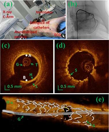
The imaging capability of the catheter was proved in vivo in a 4‑month‑old domestic porcine who was anesthetized first for the in vivo intracoronary imaging as shown in Fig. 1(a) and 1(b). The intracoronary images captured by this spacer-removed catheter are shown in Fig. 1(c) to 1(e), where Fig. 1(c) and 1(d) are the cross-section images of the blood vessel without and with stent attachment, respectively. Fig. 1(e) is cutaway view of the 3D image of a segment of coronary artery with stent and guide wire.
These results prove the effectiveness of this type of catheter in intravascular OCT imaging application. Currently, this design is being put into mass production in Shenzhen Vivolight Medical Device & Technology company, Shenzhen, China.
More information: Jiqiang Kang et al, Pencil-beam scanning catheter for intracoronary optical coherence tomography, Opto-Electronic Advances (2022). DOI: 10.29026/oea.2022.200050


