This article has been reviewed according to Science X's editorial process and policies. Editors have highlighted the following attributes while ensuring the content's credibility:
fact-checked
peer-reviewed publication
trusted source
proofread
Modeling the turtle brain provides insights into routing activity in the visual cortex

A new study by researchers at the Max Planck Institute for Brain Research uses computer simulations to explore how patterns of spikes propagate in neuronal networks constrained by experimental data from the turtle visual cortex.
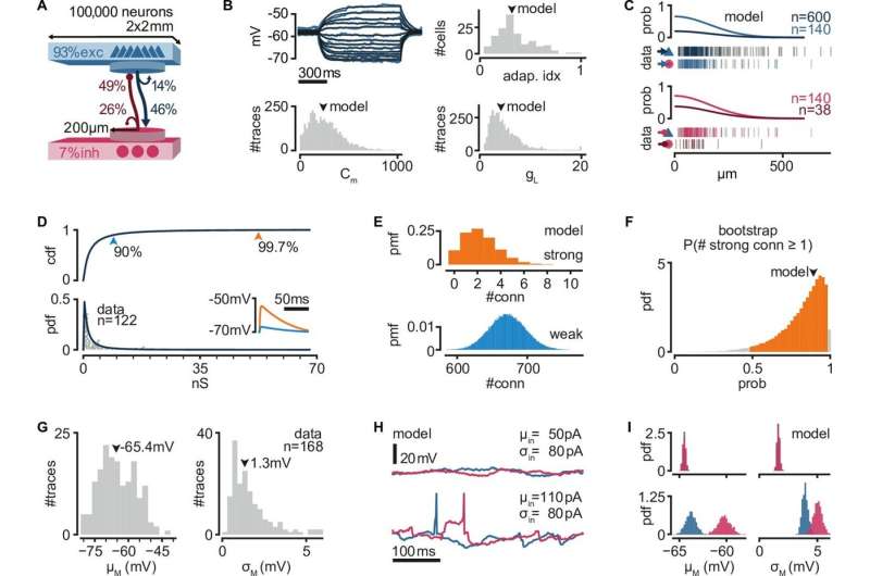
The researchers found that rare but strong connections in the network could promote the reliability of propagation, providing a substrate to easily halt or promote propagation, resulting in a highly reliable system to route activity within these networks. The research provides insight into how neurons in the brain communicate with one another and how this communication can be reliably controlled.
How reliably can networks of interconnected neurons propagate activity? Two factors argue "very unreliably": neurons receive input from many other neurons in the network through weak connections and neuronal responses vary significantly under multiple presentations of the same stimulus.
Nonetheless, repeatable patterns of activity have been measured in the brains of a variety of species, including mice, humans, birds, and turtles. These patterns involve the activation of the same network of neurons in the same temporal order, suggesting that the propagation of activity in the networks is somehow more reliable than previously believed.
Previous experiments in the laboratory of Professor Gilles Laurent, Director at the Max Planck Institute for Brain Research, had already challenged these expectations, showing that the activation (a spike) from even a single neuron in the turtle cortex can trigger patterns of activity in the network. However, the network mechanisms behind the reliable propagation of these spikes remain unclear.
Juan Luis Riquelme, a Ph.D. student working with Julijana Gjorgjieva, research group leader at the Max Planck Institute for Brain Research and Professor at TU Munich, used computer simulations to investigate the mechanism underlying activity propagation in neuronal network models.
Their study reveals the network connectivity supporting the generation of sequential neural responses by a single spike. The networks are constrained by recordings from the turtle cortex, explaining how single spikes might be able to have such an outsized effect on broad-scale neural activity.
The researchers found that rare but strong connections in the recurrent network could promote the reliability of propagation despite the presence of noisy activity from other interconnected neurons. The team observed that having a few strong connections can easily halt or promote propagation, resulting in a highly reliable system to route activity within the networks. In contrast, having many weak connections makes propagation flexible.
This routing could be context-dependent, with either external inputs or ongoing network activity determining the final path of propagation within the network. Importantly, the researchers found that these activity patterns can be separated into individual modules which can be independently activated and combined.
This suggests that the brain is capable of generating a vast number of different but reliable patterns of activity in response to different stimuli. These findings have important implications for our understanding of how the brain controls the flow of information and how it processes different stimuli.
Overall, this new computational study provides insights into how neurons in the brain communicate with one another and how this communication can be reliably controlled. As turtles are closely related to ancestral amniotes, understanding reliable propagation in their simpler cortices may provide insight into the fundamental operation of networks of neurons across multiple species.
Further research in this area could lead to insights into how the brain processes information and inform new development on artificial networks.
The study is published in the journal eLife.
More information: Juan Luis Riquelme et al, Single spikes drive sequential propagation and routing of activity in a cortical network, eLife (2023). DOI: 10.7554/eLife.79928


