This article has been reviewed according to Science X's editorial process and policies. Editors have highlighted the following attributes while ensuring the content's credibility:
fact-checked
peer-reviewed publication
trusted source
proofread
Creating a rat model for testing cell therapies in Duchenne muscular dystrophy

Duchenne muscular dystrophy (DMD) is a severe and progressive muscle disorder in which muscle degeneration and necrosis occur due to the absence of a protein called dystrophin. There is no cure for DMD, and as the disease progresses, respiratory and cardiac functions deteriorate, potentially leading to fatal respiratory or heart failure.
CiRA Associate Professor Hidetoshi Sakurai and his group previously succeeded in inducing human-iPS cells into skeletal muscle stem cells with high regenerative abilities. Next, as a proof of principle experiment for DMD cell transplantation therapy, a model animal was necessary to demonstrate the effectiveness of cell transplants.
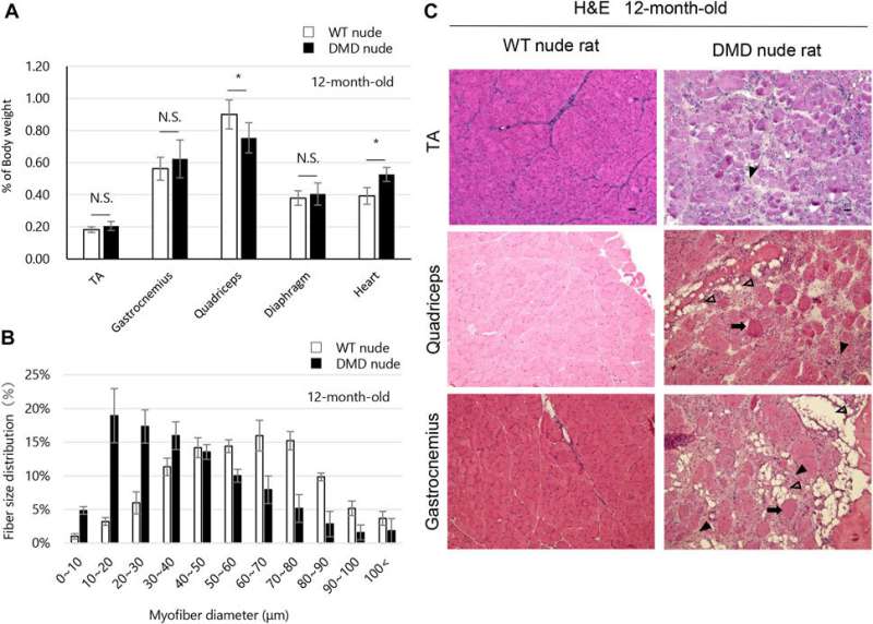
To accomplish this, the researchers created an immunodeficient DMD rat model that enables the transplantation of human cells and conducted pathological, biochemical, and motor function analyses. The results of this study were published online in Frontiers in Physiology on April 10, 2023.
This new immunodeficient DMD rat model exhibits severe symptoms similar to those observed in DMD patients. The team also reported the successful engraftment of human immortalized skeletal muscle progenitor cells into the tibialis anterior muscle of immunodeficient DMD rats, which was unsuccessful in immunocompetent DMD rats. This new model could thus be used in preclinical evaluations of cell-based therapies, allowing for more accurate measurements of motor function and biochemical tests to assess therapeutic effects.
These results will contribute to the development and early practical application of effective cell-based therapies for DMD.
More information: Masae Sato et al, A new immunodeficient Duchenne muscular dystrophy rat model to evaluate engraftment after human cell transplantation, Frontiers in Physiology (2023). DOI: 10.3389/fphys.2023.1094359


