This article has been reviewed according to Science X's editorial process and policies. Editors have highlighted the following attributes while ensuring the content's credibility:
fact-checked
trusted source
proofread
Fighting racial bias in next-gen breast cancer screening

Breast cancer is one of the most common and deadly types of cancer, and the best outcomes stem from early detection. But some screening techniques may be less effective for people with darker skin.
Seonyeong Park of the University of Illinois Urbana-Champaign will discuss experiments to measure this bias in her talk, "Virtual imaging trials to investigate impact of skin color on three-dimensional optoacoustic tomography of the breast." The presentation is part of the 184th Meeting of the Acoustical Society of America.
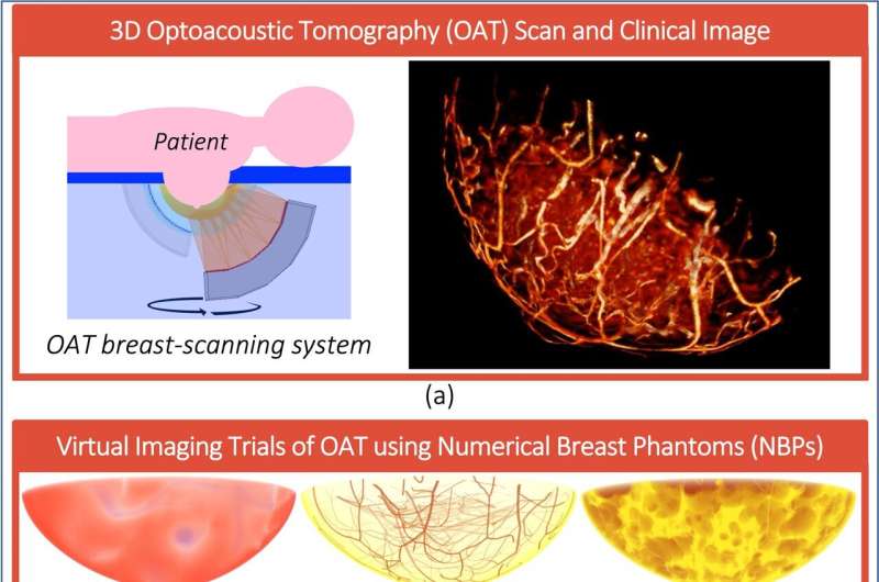
Current standard screening for breast cancer is done with X-ray mammography, which can be uncomfortable and is less effective on dense breast tissue. An alternative, optoacoustic tomography, uses laser light to induce sound vibrations in breast tissue. The vibrations can be measured and analyzed to spot tumors. This method is safe and effective and does not require compression during imaging.
The technology that underlies OAT imaging is not new; it has been used in pulse oximetry for decades. Concerns about its interaction with darker skin have existed for almost as long.
"In 1990, a study found that pulse oximetry was about 2.5 times less accurate in patients with dark skin," said Park. "Recently, an article suggested that unreliable measurements from pulse oximeters may have contributed to increased mortality rates in Black patients during the COVID-19 pandemic."
With OAT emerging as an effective breast cancer screening method, Park and her team, led by professors Mark Anastasio at UIUC and Umberto Villa at the University of Texas at Austin, collaborating with professor Alexander Oraevsky of TomoWave Laboratories, Inc. in Houston, wanted to determine if this same bias was present. Rather than navigate the cost and ethics issues surrounding human test subjects, the team instead simulated a range of skin colors and tumor locations.
"By using an ensemble of realistic numerical breast phantoms, i.e., digital breasts, the evaluation can be conducted rapidly and cost-effectively," said Park.
The results confirmed that tumors could be harder to locate in individuals with darker skin depending on the design of the OAT imager and the location of the tumor. Fortunately, a virtual framework developed by Park allows for more comprehensive investigations and can serve as a tool for evaluating and optimizing new OAT imaging systems in their early stages of development.
"To improve detectability in dark skin, the laser power should be increased," said Park. "It is recommended that skin color-dependent detectability should be evaluated when designing new OAT breast imagers. Our team is actively conducting in-depth investigations utilizing our virtual framework to propose effective strategies for designing imaging systems that can help mitigate racial bias in OAT breast imaging."
More information: Main meeting website: https://acousticalsociety.org/asa-meetings/
Technical program: https://eppro02.ativ.me/web/planner.php?id=ASASPRING23&proof=true


