This article has been reviewed according to Science X's editorial process and policies. Editors have highlighted the following attributes while ensuring the content's credibility:
fact-checked
trusted source
proofread
Non-invasive tissue oxygen imaging system for improved medical diagnosis and treatment

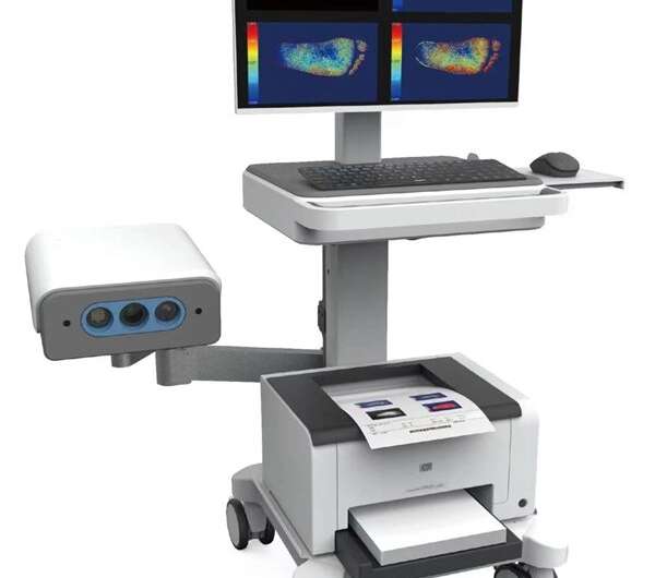
Researchers from the the Hefei Institutes of Physical Science of the Chinese Academy of Sciences have developed a new spectral imaging device called the Tissue Oxygen Imaging System (TOX).
This device enables rapid and non-invasive detection of tissue oxygen levels over a large area of at least 15 cm × 25 cm. It has recently received the official medical device registration certificate from the National Medical Products Administration of China.
Tissue oxygenation is an important indicator of tissue health and can aid in the diagnosis and treatment of various diseases. The TOX uses spatial frequency domain spectral imaging, a novel technique, to accurately assess tissue perfusion. It has shown particular efficacy in the diagnosis of diabetic foot, peripheral vascular disease, burns, and skin flap transplantation.
The TOX projects structured light of different wavelengths, modulation frequencies, and modulation phases onto the target tissue. By imaging the diffuse reflection of the tissue and using the "three-phase demodulation method," it was possible to determine how the optical parameters of the tissue are distributed in space.
The Lambert-Beer rule was used to determine how much oxygen was in the blood by looking at the concentrations of oxyhemoglobin and deoxyhemoglobin. The measurements and images helped doctors figure out how well tissue oxygenation was working.
Clinical research with 117 subjects confirmed the clinical value of this technology. The Per Protocol Set (PPS) was used as the primary dataset to assess efficacy, while the Full Analysis Set served an ancillary dataset. Statistical analysis showed minimal discrepancies between the FAS and PPS datasets, indicating good detection accuracy of the TOX.
The research team has devoted significant efforts to biomedical optics research, specializing in the extraction of weak spectral signals from tissue, recovery of intrinsic fluorescence spectra, and analysis of multispectral data from tissues.
It is expected that this technology will find more applications and innovative breakthroughs in the future, contributing to healthy aging.
Compared with traditional monitoring methods, this novel tissue oxygen detection technology offers advantages such as high accuracy, fast detection speed, and broad clinical applicability.


