This article has been reviewed according to Science X's editorial process and policies. Editors have highlighted the following attributes while ensuring the content's credibility:
fact-checked
peer-reviewed publication
proofread
New evidence for sub-network specializations within the default mode network of brain activation and self-perception

Recent advancements in neuroscience have unveiled new insights into the neural processes responsible for self-referential cognition. This research has brought particular attention to a critical neural network known as the default mode network (DMN), comprising brain regions such as the medial prefrontal cortex (mPFC), posterior cingulate cortex, temporoparietal junction (TPJ), and both lateral and medial temporal lobes.
Central to self-related processing, is the information associated with one's own body, bodily sensations, and bodily motion. Techniques like progressive muscle relaxation (PMR), which involve intentional and sequential movement of different body parts, can be used as meditative tools to enhance self-awareness. PMR is a specific protocol involving consecutive tensing and releasing of different muscle groups, promoting an increased sense of the physical self, as well as fostering relaxation.
The new study from researchers at Reichman University in Herzliya Israel, explored how PMR affects different regions of the DMN, integrating the multisensory processing of bodily signals with self-related processes. As these processes can be considered to be self-referential, DMN activation would be expected, especially with movement of body parts like the face or trunk.
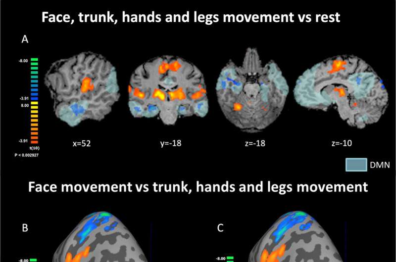
By employing functional magnetic resonance imaging (fMRI), as well as resting state functional connectivity analysis, the team of researchers investigated a novel approach of DMN activation during PMR, focusing on differential activation patterns resulting from the movement of different body parts.
Their results showed that movement of several body parts led to deactivation in several nodes of the DMN, the temporal poles, hippocampus, medial prefrontal cortex (mPFC), and posterior cingulate cortex. Notably, they found that in some of these same areas, facial movement specifically induced an inverted and selective positive BOLD pattern.
Areas in the temporal poles selective for face movement showed functional connectivity with the hippocampus and mPFC as well as with the nucleus accumbens, part of the neural circuitry for reward.
The areas activated by facial movements also exhibited strong connectivity among themselves but did not similarly connect with other regions of the DMN.
Notably, the temporal pole showed the most robust activation, especially in comparison to activations in response to movements of other body parts.
The study adds to a growing body of research highlighting the importance of facial recognition and movement in our cognitive and social development. Past research has shown that people are quicker at recognizing their own faces and making judgments about them compared to others' faces. In demonstrating how facial movements uniquely impact brain activity, the researchers further underscore the value of facial movement in our understanding of ourselves and potentially even our own emotional states.
The researchers' findings may be leveraged for therapeutic interventions, as body-related motor processes modulate the activity of nodes of the DMN, and facial movement may affect the nucleus accumbens. The summation of these areas represent regions associated with a wide range of processes, including the core self, self-referential activity, theory of mind, affective regulation, decision-making, feelings of pleasure and satisfaction, mood, and motivation.
Furthermore, they are involved in conditions such as mood disorders, anxiety, and addictive behavior. Therefore, beyond direct emotional and psychological interventions these processes can also be accessed and modulated through various body-based interventions.
Overall, the study suggests that the unique role of facial movements in brain activation may be further explored for targeted therapies that address emotional and psychological issues, and offer new avenues in treating physical health conditions.
The researchers illuminate the unique relationship between facial movements and brain activity, offering fresh insights into self-perception and mental health. This breakthrough could pave the way for new research and therapies focusing on the intricate connection between our body movements and mental states.
The research is published in the journal Restorative Neurology and Neuroscience.
More information: Inbal Linchevski et al, Integrating mind and body: Investigating differential activation of nodes of the default mode network, Restorative Neurology and Neuroscience (2023). DOI: 10.3233/RNN-231334


