This article has been reviewed according to Science X's editorial process and policies. Editors have highlighted the following attributes while ensuring the content's credibility:
fact-checked
proofread
Deciphering reproductive aging in women using a NOD/SCID mouse model

A new research paper titled "Deciphering reproductive aging in women using a NOD/SCID mouse model for distinct physiological ovarian phenotypes has been published in Aging.
Female fertility is negatively correlated with age, with noticeable declines in oocyte quantity and quality until menopause. To understand this physiological process and evaluate human approaches for treating age-related infertility, preclinical studies in appropriate animal models are needed.
In this new study, researchers María Marchante, Noelia Ramirez-Martin, Anna Buigues, Jessica Martinez, Nuria Pellicer, Antonio Pellicer, and Sonia Herraiz from IVIRMA, University of Valencia and Instituto Investigación Sanitaria La Fe aimed to characterize an immunodeficient physiological aging mouse model displaying ovarian characteristics of different stages during women's reproductive life.
"The main purpose of our study was to establish a physiological ovarian aging mouse model that could be employed to evaluate potential therapeutic interventions derived from human origin," the researchers explain.
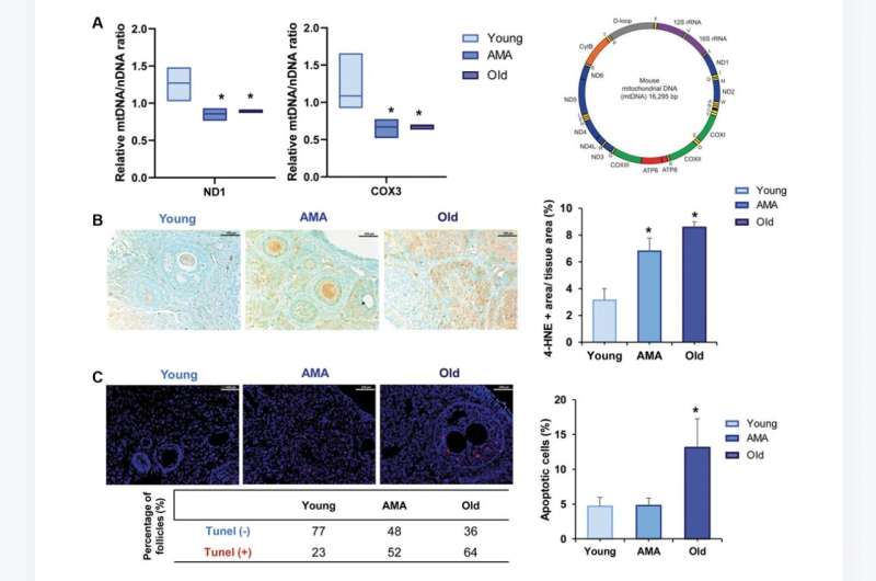
NOD/SCID mice of different ages (8-, 28-, and 36–40-weeks old) were employed to mimic ovarian phenotypes of young, advanced maternal age (AMA), and old women (~18–20, ~36–38, and >45 years old, respectively). Mice were stimulated, mated, and sacrificed to recover oocytes and embryos. Then, ovarian reserve, follicular growth, ovarian stroma, mitochondrial dysfunction, and proteomic profiles were assessed. Age-matched C57BL/6 mice were employed to cross-validate the reproductive outcomes.
The quantity and quality of oocytes were decreased in AMA and old mice. These age-related effects associated spindle and chromosome abnormalities, along with decreased developmental competence to blastocyst stage. Old mice had fewer follicles, impaired follicle activation and growth, an ovarian stroma inconducive to growth, and increased mitochondrial dysfunctions. Proteomic analysis corroborated these histological findings. Based on that, NOD/SCID mice can be used to model different ovarian aging phenotypes and potentially test human anti-aging treatments.
The researchers write, "In summary, in this study we characterized the quality of the ovarian microenvironment and reproductive outcomes of an immunodeficient murine model of physiological ovarian aging by evaluating fertility outcomes, ovarian reserve and stroma, mitochondrial dysfunctions, and the ovarian proteome at different stages. This model adequately mimicked the characteristics of the reproductive stages in women, without external agents compromising folliculogenesis, or disrupting molecular mechanisms and ovarian function, which could mask the processes of physiological aging."
More information: María Marchante et al, Deciphering reproductive aging in women using a NOD/SCID mouse model for distinct physiological ovarian phenotypes, Aging (2023). DOI: 10.18632/aging.205086


