This article has been reviewed according to Science X's editorial process and policies. Editors have highlighted the following attributes while ensuring the content's credibility:
fact-checked
peer-reviewed publication
trusted source
proofread
Researchers determine underlying mechanisms of inherited disorder that causes bone marrow failure

An international study led by researchers from Children's Hospital of Philadelphia (CHOP) has discovered an important biological cause of Fanconi anemia, a rare inherited disorder that almost universally leads to bone marrow failure. The researchers also confirmed that a readily available bile acid may help correct some of these biological issues and provide more options for potential treatment. The findings were recently published by Nature Communications.
Fanconi anemia was discovered nearly a century ago. Over the course of decades of study, researchers had previously linked Fanconi anemia to DNA damage that impairs the function of hematopoietic stem cells (HSCs), which are responsible for lifelong function and renewal of blood cells.
DNA damage leads to cell death and eventually gives way to frank bone marrow failure. Since stem cells are impacted, a stem cell transplant is the only treatment currently available for Fanconi anemia. However, not all patients have matching donors, and those who do can still experience side effects from the treatment.
Prior research by the group revealed that the loss of stem cells that leads to Fanconi anemia begin before birth, making the underlying biology behind this condition very difficult to study. Using mouse models and a combination of other techniques, researchers were able to determine the events before birth that eventually accelerated bone marrow failure by early school age in these patients.
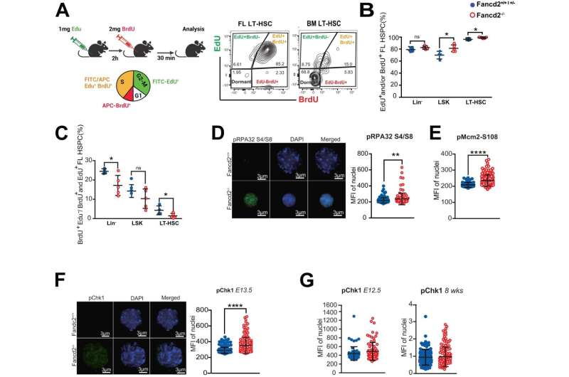
"Fanconi anemia is thought to be a DNA disorder, and those who have been researching it have focused solely on this aspect," said senior author Peter Kurre, MD, Director of the Pediatric Comprehensive Bone Marrow Failure Center at CHOP. "What our research demonstrates for the first time is that Fanconi anemia is caused by an accumulation of misfolded proteins that preclude proper cell cycle progression and stem cell expansion, which in turn leads to rapid bone marrow failure after birth."
The study found that increased protein synthesis rates in fetal HSCs occur at the onset of Fanconi anemia. The proteins are misfolded in fetal liver HSCs, which leads to stress on the endoplasmic reticulum, a network of tubules in our cells responsible for producing healthy proteins needed for a variety of functions.
This accumulation of misfolded proteins confers cellular stress signals that slow down cell cycle progression and the emergence of a sufficient number of stem cells, causing rapid bone marrow failure early in life.
With this new knowledge of the mechanisms behind Fanconi anemia, the researchers then used tauroursodeoxycholic acid (TUDCA), a bile salt that has been studied for use in preventing and treating gallstones and helping to reduce the side effects associated with certain inflammatory disorders. In the HSC of the Fanconi anemia animal model, TUDCA restored proper protein folding.
"When TDUCA was given, we were able to confirm the restoration of folding as well as improved function of the stem cells," said Narasaiah Kovuru, Ph.D., a postdoctoral researcher in the Kurre Laboratory and first author of the study.
With this combination of information, the researchers determined that the degradation of protein homeostasis—the proper regulation of proteins in these cells—is driven by excess inflammatory activity in HSCS within the fetal liver, and dampening this activity helps restore HSCs to their normal numbers.
"Regulating protein production, folding, and degradation properly is like the story of Goldilocks and the Three Bears, in that too much or too little protein is a problem, and the levels need to be just right for proper function," Kurre said. "This study reveals the direction our research should go next to determine what mechanisms are at play that alter these protein levels and what alternate treatment options we might be able to develop for patients for whom stem cell transplantation is not an option."
More information: Narasaiah Kovuru et al, Deregulated protein homeostasis constrains fetal hematopoietic stem cell pool expansion in Fanconi anemia, Nature Communications (2024). DOI: 10.1038/s41467-024-46159-1


