This article has been reviewed according to Science X's editorial process and policies. Editors have highlighted the following attributes while ensuring the content's credibility:
fact-checked
proofread
Altered brain morphology and functional connectivity in postmenopausal women

A new research paper titled "Altered brain morphology and functional connectivity in postmenopausal women: automatic segmentation of whole-brain and thalamic subnuclei and resting-state fMRI" has been published in Aging.
The transition to menopause is associated with various physiological changes, including alterations in brain structure and function. However, menopause-related structural and functional changes are poorly understood.
In this new study, researchers Gwang-Won Kim, Kwangsung Park, Yun-Hyeon Kim, and Gwang-Woo Jeong from Chonnam National University not only compared the brain volume changes between premenopausal and postmenopausal women, but also evaluated the functional connectivity between the targeted brain regions associated with structural atrophy in postmenopausal women.
"To the best of our knowledge, no comparative neuroimaging study on alterations in the brain volume and functional connectivity, especially focusing on the thalamic subnuclei in premenopausal vs. postmenopausal women, has been reported," the researchers state.
Each of the study's 21 premenopausal and postmenopausal women underwent magnetic resonance imaging (MRI). T1-weighted MRI and resting-state functional MRI data were used to compare the brain volume and seed-based functional connectivity, respectively. In statistical analysis, multivariate analysis of variance--with age and whole brain volume as covariates--was used to evaluate surface areas and subcortical volumes between the two groups.
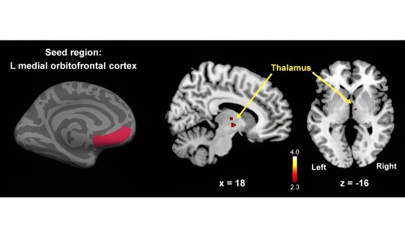
Postmenopausal women showed significantly smaller cortical surface, especially in the left medial orbitofrontal cortex (mOFC), right superior temporal cortex, and right lateral orbitofrontal cortex, compared to premenopausal women (p < 0.05, Bonferroni-corrected) as well as significantly decreased functional connectivity between the left mOFC and the right thalamus was observed (p < 0.005, Monte-Carlo corrected).
Although postmenopausal women did not show volume atrophy in the right thalamus, the volume of the right pulvinar anterior, which is one of the distinguished thalamic subnuclei, was significantly decreased (p < 0.05, Bonferroni-corrected).
"Postmenopausal women showed significantly lower left mOFC, right lOFC, and right STC surface areas, reduced right PuA volume, and decreased left mOFC-right thalamus functional connectivity compared to premenopausal women. If replicated in an independent sample, these findings will be helpful for understanding the effects of menopause on the altered brain volume and functional connectivity in postmenopausal women," the researchers conclude.
More information: Gwang-Won Kim et al, Altered brain morphology and functional connectivity in postmenopausal women: automatic segmentation of whole-brain and thalamic subnuclei and resting-state fMRI, Aging (2024). DOI: 10.18632/aging.205662


