This article has been reviewed according to Science X's editorial process and policies. Editors have highlighted the following attributes while ensuring the content's credibility:
fact-checked
peer-reviewed publication
trusted source
proofread
International collaboration produces a comprehensive atlas of human skeletal muscle aging

In a world with rapidly aging societies, there's a need for a detailed understanding of the cause and progression of diseases associated with aging. Skeletal muscle is the key motor system in the human body and plays a pivotal role in body metabolic regulation. With increased age, particularly in individuals over 80 years old, skeletal muscles suffer from sarcopenia, a progressive loss of muscle mass and function.
Sarcopenia not only increases the individual's disability but also plays a role in the rapid decline of general functions in the elderly, making them frailer. The underlying mechanisms are not well understood. Until now, the biological basis of sarcopenia at single-cell level had not been investigated systematically.
Scientific research teams from Pompeu Fabra University (UPF) in Barcelona (Spain), Altos Labs in San Diego (U.S.A.), Valencia University/INCLIVA and Hospital Arnau de Vilanova in Valencia (Spain), BGI-Research, The First Affiliated Hospital of Guangdong Pharmaceutical University, Guangzhou Institutes of Biomedicine and Health (Chinese Academy of Sciences), and other institutions, analyzed the gene expression and epigenetic status of 387,000 individual cells in lower limb muscle biopsies from 31 individuals of different genders, ages and regional backgrounds.
With this data, they have outlined the most comprehensive single cell atlas of aging human skeletal muscle to date. This breakthrough study, "Multimodal cell atlas of the ageing human skeletal muscle," is published in Nature.
This international collaborative effort was led by Dr. Pura Muñoz-Cánoves, an ICREA research professor at the Medicine and Life Sciences Department at UPF in Barcelona, and now a principal investigator in the Altos Labs San Diego Institute of Science, and Dr. Miguel. A. Esteban at BGI-Research in Shenzhen.
"As the most exhaustive atlas of human aging muscle at the single cell level to date, this study will be a reference for the fields of both aging and sarcopenia and frailty," said Dr. Pura Muñoz-Cánoves.
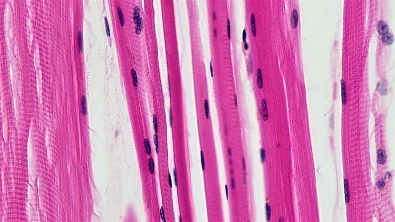
Human skeletal muscle is largely made up of muscle fibers (myofibers), of which there are two types.
Type 1 muscle fibers are primarily involved in endurance physical activity, such as long-distance running or cycling. They are characterized by a slow muscle contraction speed, high aerobic metabolism, and rich mitochondria activity.
Type 2 muscle fibers are important in physical activities that require sudden bursts of power such as jumping, sprinting and weightlifting. They have faster muscle contraction rates, are more prone to fatigue, and rely mainly on anaerobic metabolism to produce energy.
This work describes how skeletal muscle cell populations, including both the individual nuclei in multinucleated fibers and in conventional mononucleated cells, change with aging, as well as the multi-cell networks underlying these changes. By comparing this data with genetic data, the team was also able to identify key elements that predict susceptibility to sarcopenia.
The researchers found that as humans age, type 2 muscle fibers deteriorate steadily during the aging process, while type 1 muscle fibers remain relatively stable and better tolerate the stress of aging. During the aging process, cell metabolism is also affected. While type 1 fibers become more glycolytic, type 1 muscle fibers increase oxidation. Importantly, novel pro-regenerative and pro-degenerative myofiber subtypes emerge upon aging. These new populations may be instrumental in inducing the degenerating cascade of aging muscle and are likely targets for intervention.
Muscles can repair themselves. This is mostly done by muscle stem cells that, upon injury, begin to proliferate and differentiate into muscle, fusing with each other or with existing muscle fibers to repair damaged muscle. The researchers found that these stem cells exit the quiescent state in aging muscles and enter a premature priming state, resulting in reduced regeneration capacity.
Meanwhile, during aging, endothelial cells also undergo changes with increased pro-inflammatory and chemotactic signals, while immune cells increase in number and initiate inflammatory programs. These changes make muscles more susceptible to deterioration in response to injury and may promote systemic inflammation and accelerate the decline of overall physical function in older people.
In addition, through cross-comparison with genetic data, the researchers identified cell-type-specific sites in chromatin, the mixture of DNA and proteins that forms chromosomes in human cells, associated with susceptibility to sarcopenia. These findings provide researchers with potential new targets for the future diagnosis and treatment of sarcopenia.
Dr. Miguel A. Esteban, one of the two co-corresponding authors of this study, said, "Our joint scientific research provides a new perspective to understand human skeletal muscle aging and an exciting scientific basis for the development of preventative and therapeutic strategies."
"This atlas is the product of an international collaboration and the development of massively parallel single-cell profiling technologies," said Dr. Yiwei Lai, first author of the study and a member of the Chinese team.
"Our single nucleus expression analysis has allowed the possibility of studying cell populations that could not be characterized by conventional studies, such as myonuclei from the multinucleated skeletal muscle fibers," said Ignacio Ramírez-Pardo, one of the co-first authors of the study, from UPF and Altos Labs.
Other relevant contributors to the study, Dr. Joan Isern, Eusebio Perdiguero and Antonio Serrano from Altos Labs and UPF teams, coincided in adding that "it will be important to compare this human muscle aging atlas with previous cell atlases from non-human primates and from other species, as it will help establish interspecies adaptive comparisons and predict disease susceptibility."
Doctors Mari Carmen Gómez-Cabrera and Julio Doménech-Fernández (from Valencia University/INCLIVA and Hospital Arnau de Vilanova in Valencia, respectively) highlighted that "this atlas will also be an important reference for future studies in patients with neuromuscular diseases."
"We hope that this will be the basis of many subsequent investigations to slow down or even block sarcopenia, frailty, and muscle deterioration in older people, promoting healthier body aging for longer and increasing longevity," said Dr. Pura Muñoz-Cánoves.
This study demonstrates the importance of international cooperation and multidisciplinary teamwork in addressing critical scientific challenges.
By further expanding the sample size and using muscle samples from other parts of the body in different contexts, the research team aims to build a more comprehensive atlas to improve the understanding of muscle function and muscle aging and offer optimism in tackling the challenges faced by aging societies.
More information: Pura Muñoz-Cánoves, Multimodal cell atlas of the ageing human skeletal muscle, Nature (2024). DOI: 10.1038/s41586-024-07348-6. www.nature.com/articles/s41586-024-07348-6


