This article has been reviewed according to Science X's editorial process and policies. Editors have highlighted the following attributes while ensuring the content's credibility:
fact-checked
peer-reviewed publication
proofread
New molecular sensor enables fluorescence imaging for assessing sarcoma severity

Researchers at Korea University College of Medicine have identified a new candidate marker for determining the severity and metastasis of sarcoma and developed a molecular sensor that enables fluorescence imaging targeting this marker.
The advance has been published as the cover article in Angewandte Chemie International Edition.
Sarcoma is a broad group of cancers that originates in the soft tissues. Due to its heterogenous nature, it has been challenging to quantitatively assess the severity and metastasis of sarcomas in clinical pathology, making diagnosis and prognosis monitoring difficult.
Furthermore, conventional cancer stem cells (CSCs) markers often exhibit an overexpression in heterogeneous malignancies, complicating the identification and isolation of CSCs within tumor cells.
The research team, led by Professors Jun-Seok Lee (Department of Pharmacology) and Woo Young Jang (Department of Orthopedic Surgery), discovered a correlation between the expression of the conventional CSC marker (CD44) and the Prostaglandin synthesis network. They found that Cyclooxygenase (COX) expression exhibited statistical specificity across different sarcomas.
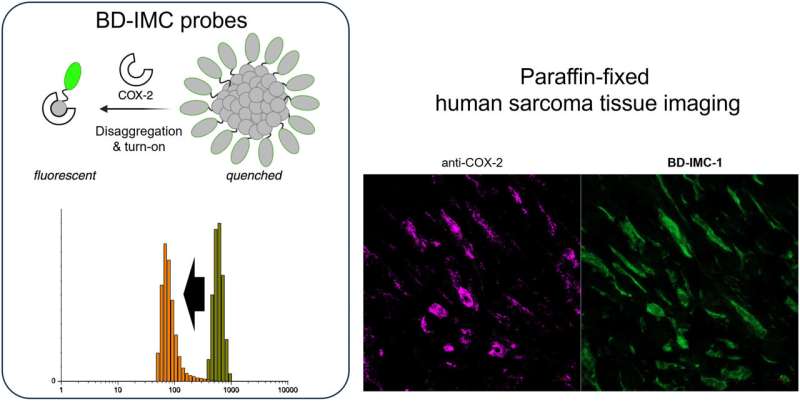
Based on these findings, the researchers designed two fluorescent probes (BD-IMC-1, BD-IMC-2) that target COX enzymes and activate fluorescence upon disaggregation. This innovative approach enables the visualization of CSCs within sarcoma tissues.
Specifically, they linked BODIPY fluorescent molecules to the COX inhibitor indomethacin, designing molecules that induce self-aggregation of nanostructures and remain in a quenched fluorescent state in aqueous solutions. These molecules exhibit fluorescence sensitively only when bound to COX enzymes, functioning as chemosensors. By utilizing COX inhibitors and fluorescent structures to disaggregate fluorescent molecules, they designed an imaging sensor that activates fluorescence.
In the process, they also identified new candidate markers, suggesting the necessity for further systematic research on the correlation between COX expression and CSC expression within sarcoma tissues.
Previously, imaging molecules targeting COX enzymes induced changes in fluorescence characteristics at the single-molecule level to visualize COX enzymes. However, this study is the first to report imaging target proteins in fixed clinical samples based on the fluorescence characteristics of structural changes in fluorescent multicomplexes.
Professor Jun-Seok Lee from the Department of Pharmacology stated, "The newly developed fluorescent molecular sensor does not rely on changes in fluorescence characteristics at the single-molecule level but utilizes the self-aggregated state and characteristics of multiple molecules, making it effective in complex samples such as biological tissues."
He added, "This research represents a new strategy for developing imaging sensors for various biological targets, contributing to the development of imaging-based diagnostic and prognostic monitoring techniques for sarcoma."
More information: Kyung Tae Hong et al, Disaggregation‐Activated pan‐COX Imaging Agents for Human Soft tissue Sarcoma, Angewandte Chemie International Edition (2024). DOI: 10.1002/anie.202405525


