January 29, 2014 report
Histological sectioning and digital 3D reconstruction of H.M's brain detailed

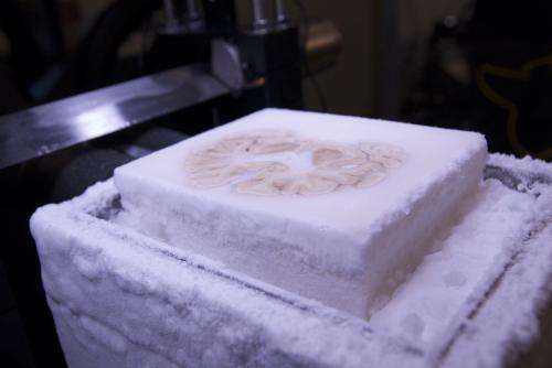
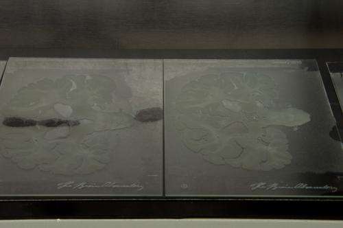
(Medical Xpress)—The team of researches that sliced up the brain of famous neurological patient H.M has published a postmortem examination of their work in the journal Nature Communications. In their paper, the team describes the techniques they used to cut the brain into 2401 slices, some of which were dyed for cell study, and how each slice was photographed for creating a 3D replica of the entire brain.
H.M. as he's been known in neurological journals for several decades, has been revealed to be Henry Molaison, a man who at age 27 agreed to allow neurosurgeon William Scoville to cut out part of his brain (back in 1953) as a last-ditch effort to relieve chronic seizures due to epilepsy. That operation, which did indeed reduce the seizures, also had a surprising side-effect—Molaison's brain was no longer able to create memories that would last more than a few minutes.
Prior to the surgery on Molaison, scientists believed that memory formation was spread throughout the brain. In removing large parts of the temporal lobe, which included the hippocampus, Scoville inadvertently proved that certain parts of the brain are much more involved in creating and retaining memories than others.
Over the course of his lifetime, Molaison underwent several MRI's which helped neuroscientists better understand what went on with his brain. He also participated in a large variety of other tests with other researchers with the goal of better understanding the role of different parts of the brain in memory processing. As a result, Molaison has become the most cited patient in the history of neuroscience.

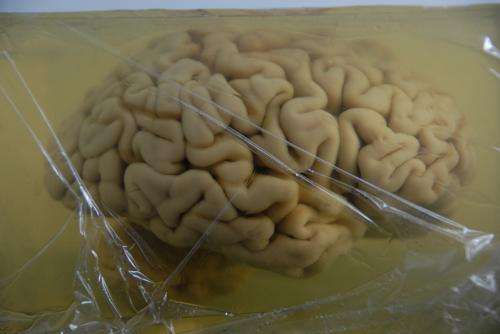
In their paper describing their postmortem examination, the team described how they preserved Molaison's brain after he died in 2008 by encasing it in gelatin and freezing it. The brain was placed in a device that was able shave off paper-thin slices, each of which were digitally photographed and subsequently compiled to build a faithful 3D representation of the original brain.

In analyzing the result, the team noted that less of the hippocamus was removed in the surgical procedure to cure Molaison of his seizures than was first believed. They also found a previously unknown lesion that had formed in another part of his brain. More noteworthy perhaps is that the team filmed the postmortem and along with the video that was created, they have posted online the 3D brain image created, inked slides and other views of the brain taken before the postmortem, for anyone who wishes to view them.
More information: Read also: 3 Questions: Suzanne Corkin on new study of neuroscience's most famous patient
Paper: Postmortem examination of patient H.M.'s brain based on histological sectioning and digital 3D reconstruction, Nature Communications 5, Article number: 3122 DOI: 10.1038/ncomms4122
Abstract
Modern scientific knowledge of how memory functions are organized in the human brain originated from the case of Henry G. Molaison (H.M.), an epileptic patient whose amnesia ensued unexpectedly following a bilateral surgical ablation of medial temporal lobe structures, including the hippocampus. The neuroanatomical extent of the 1953 operation could not be assessed definitively during H.M.'s life. Here we describe the results of a procedure designed to reconstruct a microscopic anatomical model of the whole brain and conduct detailed 3D measurements in the medial temporal lobe region. This approach, combined with cellular-level imaging of stained histological slices, demonstrates a significant amount of residual hippocampal tissue with distinctive cytoarchitecture. Our study also reveals diffuse pathology in the deep white matter and a small, circumscribed lesion in the left orbitofrontal cortex. The findings constitute new evidence that may help elucidate the consequences of H.M.'s operation in the context of the brain's overall pathology.
© 2014 Medical Xpress


