February 7, 2014 feature
Ending diffusion confusion: Identifying precise neural correlates of autism spectrum disorder

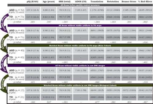
(Medical Xpress)—Autism Spectrum Disorder (ASD) refers to a group of developmental disorders (such as autism and Asperger's syndrome) characterized by impairments in the ability to communicate and interact socially. ASD has a generally well-accepted brain signature – namely, the reduced integrity of long-range white-matter fiber tracts consisting mostly of glial cells and myelinated axons that transmit signals from one region of the cerebrum to another and between the cerebrum and lower brain centers. These tracts are investigated primarily through diffusion imaging studies. (Based on the random thermal motion of molecules, diffusion imaging – specifically, Diffusion Magnetic Resonance Imaging or dMRI – enables the imaging of white matter in the brain by measuring the magnitude and orientation of water diffusion in multiple directions to calculate a three-dimensional water diffusion profile, which in the case of dense white matter tracks is highly anisotropic, or ovoid, pointing in the direction of the fiber bundle.) Recently, scientists at Massachusetts Institute of Technology and Massachusetts General Hospital assessed known white matter tracts in children with ASD by using Diffusion-Weighted Imaging (DWI-MRI), where the image contrast is determined by the Brownian (random) microscopic motion of water protons. Unlike most previous studies, however, the researchers carefully matched head motion between groups, and in doing so demonstrated that there was no evidence of widespread changes in white-matter tracts in the ASD group – rather, differences were present only in the right inferior longitudinal fasciculus (rILF), a white matter tract that connects the temporal and occipital lobes. They conclude that their data challenge the idea that widespread changes in white-matter integrity are a signature of ASD and highlight the importance of matching for data quality in future diffusion studies of ASD and other clinical disorders.
Discussing the paper that she, Prof. Nancy Kanwisher and their co-authors published in Proceedings of the National Academy of Sciences, Dr. Kami Koldewyn first tells Medical Xpress about the challenges they faced in conducting their research, beginning with their decision to target head motion artifacts as the key factor in potentially erroneous multiple white matter tract findings. "Several recent papers have shown how head motion during scanning can directly result in spurious between-group findings in functional connectivity studies," Koldewyn points out, adding that they wondered if the same might prove to be true in Diffusion Tensor Weighted Magnetic Resonance Imaging (DW-MRI) as well. "We know that head motion during DW-MRI scanning leads to a decrease in image intensity," she explains, "thus changing the very phenomenon that produces the DW-MRI contrast. This means that any difference between groups in how much they move inside the scanner could lead directly to between-group differences in DW-MRI measures. The fact that very few papers recruiting participants with ASD measured head motion, or factored it into their analyses, meant that it was possible that differences in DWI measures between groups may have been incorrectly interpreted as less robust connections in white-matter tracts when they may have simply been differences in head movement during scanning."
Another issue was precisely matching data quality between groups, which Koldewyn acknowledges is not a simple process to accomplish with great precision. "It isn't simply the misalignment between scans that needs to fixed," she explains. "Head motion in the scanner causes image artifacts that cannot easily be compensated for." This means that the type of motion can make a difference – and the researchers' more technical paper published in Neuroimage1 examining the effects of head motion on group analyses in DWI showed that all types of motion made a difference in between-group analyses. For that reason, Koldewyn tells Medical Xpress, the scientists matched groups on each of the motion measures in the current paper, rather than on a composite motion measure. "The head motion measures we used are not as precise as we might wish them to be," she acknowledges, "and there is a clear need for future innovation in better motion measurements as well as both post-scan and in-scan compensation for head motion."
Ironically, group differences in head motion can lead artifactually to just the effects most often reported – specifically, reduced fractional anisotropy (FA) in ASD white matter tracts. (Fractional anisotropy gives a value between 0 and 1, 0 being isotropic, or spherical. FA characterizes longitudinal directional diffusion, thereby reflecting white matter fiber density, axonal diameter, and myelination.) However, the scientists found that these differences were observed across the brain before matching the groups for head motion – but, critically, not afterwards. This demonstrates that head motion really does matter when looking at group differences – a point also made in the aforementioned technical paper1, in which the scientists showed that the between-group DW-MRI differences hold true not only when comparing autism and typical groups, but also when both groups include only typically developing children. "Indeed," Koldewyn stresses, "we found that head-motion differences between groups also induce differences in DW-MRI measures even when the children in the two groups are the very same children scanned twice."
Given the above, it was vital that the researchers find evidence for reduced connectivity of only the rILF fiber tract. "Many researchers, including our group, have found that children with ASD have more difficulty remembering faces than their peers," Koldewyn points out. "In fact," she illustrates, "recent research has suggested that people who have congenital prosopagnia" – a developmental condition characterized by an impaired ability to recognize faces – "have less robust structural connectivity in the right inferior longitudinal fasciculus. We thought it might be possible that children with ASD would show a similar difference in the right inferior longitudinal fasciculus, so we were particularly interested in looking at that tract between groups."
These findings made it necessary for the scientists to determine if some of the differences between their results and the previous literature could be explained by other factors, such as differences in analysis methods such as tract-based tract‐based analysis versus voxel‐based morphometry (VBM) – the two main methods of analyzing diffusion images. (Tract‐based analysis targets white matter fiber tracks to calculate FA values averaged across the tract that can be compared across groups to investigate structural connectivity. Voxel‐based morphometry statistically compares local anisotropy values for the whole brain between different subjects.)
"We decided to adopt our particular method because it has previously been shown to be robust and accurate," Koldewyn explains. "In addition, the fact that identifying the tracts in each participant has been automatized means that there is less subjectivity in the identification of the tracts and that it was more feasible to run these analyses on a large group. When we looked at our data using a voxel-wise method, we also found that head-motion made a difference in between-group analyses. For this reason, we suspect that our findings are not dependent on our analysis technique." However, she acknowledges that it remains possible that differences between their results and previous findings could be partially driven by differences in analytic techniques.
The scientists also had to evaluate whether their method, which was originally developed for adult subjects, works as well with child participants. "A previous paper explored this exact question and found that the method we used was not affected by the age of the subjects, working just as well for children as it did for adults" Koldewyn says. "For this reason, we're confident that our results are not negatively affected by the fact that our participants were children."
Finally, the team had to determine if widespread reductions in FA are present in individuals with ASD who are younger, older, or lower-functioning than those they tested. "Our own analyses found no indication that either younger or older children with ASD showed stronger differences when compared with TD children," Koldewyn points out, "suggesting that there weren't 'hidden' group differences in only one age range – but that said, we only looked at children aged 5 – 12, so it remains possible that either very young children or older adolescents or adults could show more differences."
Looking at changes in diffusion measures in individuals with low functioning, or more severe, ASD is difficult because many of these individuals cannot tolerate being scanned. (Low functioning children with autism often exhibit little or no language, some degree of intellectual impairment, little awareness of social expectations, and may show self-injurious behaviors, especially when distressed.) "Unless imaging studies opt for performing DWI scanning while participants are anesthetized – which is typically for clinical scans, not just for research purposes – such individuals cannot contribute to the picture we get of the disorder through imaging," Koldewyn explains. "This, in turn, could potentially bias the imaging picture we have of ASD as a whole. The few DWI studies that are available from studies of lower-functioning participants suggest that the DWI differences found in this group may be more related to intellectual impairment than ASD itself. However, it's clear that more research is needed before we can draw strong conclusions."
Koldewyn says that the key insight for their paper was simply considering the possibility that head-motion could be biasing our picture of structural brain differences in ASD. "We had a great team of researchers, including people that are experts in image analysis and DWI, and working in this team helped us apply excellent tools to what is actually a pretty complex question."
Among the researchers' additional analyses was an analysis intended to show that the study had sufficient statistical power by showing the expected developmental increases in the FA of fiber tracts within ASD and typical groups individually. "Much of the meat of this paper is a null finding – we didn't find large differences between the ASD and typical groups except in one tract. One difficulty in presenting null findings is that the reader is left wondering if we had enough data (enough statistical power) to find differences between groups if they really existed. One way to argue that you have enough power to see group differences is by demonstrating that you have as much data as previous studies that have found group differences. We would have liked to do this, but the details of the statistics that we would have needed to do a power analysis were not included in any paper looking at DTI differences between ASD and TD groups with analyses that were sufficiently similar to the analyses we were conducting. So we opted for a different way of showing that we had sufficient power – by demonstrating that we could find expected group differences within the same data set but comparing younger to older children rather than children with ASD to TD children. The logic of this comparison was that we could find an expected group difference in our data set using half of the data used in the main comparison, suggesting that we should have enough data in the full data set to ensure that we could find real group differences if they were there.
Looking ahead, Koldewyn says, the scientists would like to address three areas of research:
- Group differences in the right ILF will need to be replicated in an independent sample
- Being able to use DWI-MRI measures within the ILF (and perhaps other white matter tracts) to investigate relationships between structural connectivity in specific tracts and cognitive and perceptual function in people with ASD (for instance face perception/memory abilities)
- Once head motion is accounted for, DW-MRI measures across several research groups could potentially be combined and, along with cognitive and clinical data, used to help identify sub-groups within ASD
Koldewyn adds that she would like to work in collaboration with MR physicists to help develop scans that are quieter, quicker and less sensitive to motion artifacts for use in scanning children and clinical populations – and notes that other areas of research might benefit from their study. "The implications for future research are pretty simple: head motion and other data quality differences between groups must be taken into account to avoid spurious findings. This is particularly true for difficult-to-scan populations," she emphasizes, "including practically any clinical population – for example, schizophrenia, anxiety disorders and ADHD – as well as studies comparing children across development or to adults." In addition, she recently wrote a guest blog post for SFARI2 about the technical paper mentioned above1, in which she writes that motion artifacts and data quality are relevant to other many imaging techniques beyond DWI-MRI.
"We hope that other researchers will view our paper as an invitation to look back on previous DWI datasets and take head motion and data quality into account in their future DWI research." Koldewyn concludes."Doing so will be essential for future research in clinical disorders like ASD, but is also important in work looking at typical development." To this end, Anastasia Yendiki, one of the co-authors of the paper and also the person who developed the DWI-MRI toolbox the researchers used for data analysis, has made code freely available3.
More information: Differences in the right inferior longitudinal fasciculus but no general disruption of white matter tracts in children with autism spectrum disorder, Proceedings of the National Academy of Sciences Published online before print on January 21, 2014, doi:10.1073/pnas.1324037111
Related
1Spurious group differences due to head motion in a diffusion MRI study, Neuroimage 2013 Nov 21;88C:79-90, doi:10.1016/j.neuroimage.2013.11.027
2Heeding head motion's effects
3TRACULA: TRActs Constrained by UnderLying Anatomy
© 2014 Medical Xpress. All rights reserved.


