Scientists restore normal function in heart muscle cells of diabetic rats
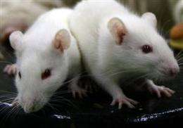
Working with heart muscle cells from diabetic rats, scientists at Johns Hopkins have located what they say is the epicenter of mischief wreaked by too much blood sugar and used a sugar-gobbling enzyme to restore normal function in the glucose-damaged cells of animal heart muscles.
In addition to much-needed insight into the process of diabetes-related heart damage, the study, described June 24 in the journal Diabetes, offers a clue to a possible treatment strategy for diabetic cardiomyopathy, a condition marked by progressive weakening of the heart muscle found in 60 percent of the 380 million people with diabetes worldwide.
"Glucose toxicity in the heart is one of the gravest complications of diabetes and one whose mechanisms are poorly understood," says lead investigator Genaro Ramirez-Correa, M.D., Ph.D., instructor of pediatric cardiology at the Johns Hopkins Children's Center and member of the Johns Hopkins Innovative Proteomics Center in Heart Failure. "Our research maps out the molecular chain of events inside the cells that is responsible for the heart muscle damage seen in diabetes."
Scientists have known for some time that hearts affected by diabetes conduct electric signals abnormally and contract aberrantly—both problems believed to stem from the cells' inability to properly respond to calcium, the chief trigger of cardiac and other muscle contraction. But how andwhy heart cells in diabetes fail to respond to contraction-fueling calcium has been a puzzle.
Ramirez-Correa says his team's findings reveal that a sugar molecule and an enzyme charged with regulating the molecule's levels inside cells appear at the root of calcium insensitivity and poor contraction. The sugar molecule, called O-GlcNAc, and one of its "handler" enzymes, OGT, get shuffled around the cell, end up in the wrong location. From there, the researchers observed, they there interfere with the work of proteins that control muscle contraction.
To glean insight into the process, the research team started out with the premise that in diabetes, cells in every organ are awash in excess glucose. The scientists homed in O-GlcNac, an end product of glucose breakdown, a process that is abnormal in people with diabetes. Using technology that analyzes proteins and visualizes their position and movement inside cells, the researchers found that O-GlcNAc levels are notably higher in cells of diabetic rats than they are in the cells of healthy rats.
Next, using a sophisticated laser microscope to obtain detailed images of the cells' interiors, researchers focused on the areas within heart cells that control muscle contraction—tiny muscle units known as sarcomeres. Scientists noticed that in diabetic heart muscle cells, O-GlcNAc and one of its chaperone enzymes, OGT, move away from the sarcomere's border and toward the middle where various proteins normally connect with each other and form chains that pull the ends of the sarcomere closer together—the essence of muscle contraction. Normally, flow of calcium into the heart cells is a call to action for muscle proteins to form a cross-bridge and initiate contraction. But when O-GlcNAc and OGT insert themselves in the middle, like struts, the researchers found, they prevented contractile proteins from cross-bridging properly with one another in response to calcium. That, the researchers say, fuels the calcium insensitivity seen in diabetic hearts and causes abnormal muscle contraction.
"When the sugar molecule and the enzyme move into the control center of muscle contraction, it's like putting a drop of molasses inside a Swiss watch," Ramirez-Correa says. "You end up disrupting a perfectly synchronized mechanism."
In further experiments, researchers used an enzyme, OGA, whose sole job is to break down O-GlcNAc. Harnessing the enzyme's appetite for O-GlcNAc, they introduced a bacterial form of this sugar gobbler into the muscle cells of diabetic hearts. The cells' ability to respond to calcium was restored and so was their normal contraction, the researchers observed. Reassuringly, Ramirez-Correa says, when scientists exposed healthy heart muscle cells to the same enzyme, nothing changed.
"Removing this molecule from the wrong location inside the heart cell is akin to getting rid of the motion-disrupting kink in a bike chain," says senior investigator Anne Murphy, M.D., a cardiologist at the Johns Hopkins Children's Center and professor of pediatrics at the Johns Hopkins University School of Medicine.
Investigators say that if their observations are confirmed in larger animals and then in humans, the findings could lead to the development of drugs that inhibit the toxic reshuffling of sugar molecules and enzymes inside heart cells. The results could also form the basis for a clinical biomarker that heralds the onset of diabetic cardiomyopathy before irreversible damage occurs.
"We know that amino acids—the building blocks of proteins—leak into the blood when heart proteins break down," Ramirez-Correa says. "When sugar molecules latch onto the heart's contractile proteins, the amino acids they release could serve as telltale footprints that signal glucose toxicity in a single blood drop from a patient with diabetes."
More information: "Removal of Abnormal Myofilament O-GlcNAcylation Restores Ca2+ Sensitivity in Diabetic Cardiac Muscle." Diabetes published ahead of print June 24, 2015, DOI: 10.2337/db14-1107


