March 25, 2014 feature
Thanks for the memories: Plant-like photopigment in the eye may play key role in human cognitive brain function

(Medical Xpress)—Light is inextricably intertwined in myriad ways with most life on Earth. In humans, for example, light stimulates alertness and cognition, improving performance and increasing wakefulness. In a recent study, scientists at University of Liège, Belgium and Stem Cell and Brain Research Institute, France demonstrated that exposure to longer wavelength light, relative to shorter wavelength, subsequently enhances the impact of light on executive brain function through the recently-discovered photopigment melanopsin, an invertebrate-like, even plant-like, photoreceptor. By combining melanopsin responses combined with functional Magnetic Resonance Imaging (fMRI) recording, the researchers concluded that since photic memory – the effects of prior light on subsequent responses to light – is typical not only of melanopsin, but of certain invertebrate and plant photopigments as well, humans may therefore have an invertebrate or plant-like machinery within the eyes that participates to regulate cognition. Moreover, they state that their findings may explain a type of long-term adaptation to previous lighting conditions known as the previous light history effect, and support the design of cognitive performance-optimizing lighting systems.
Dr. Howard M. Cooper and Prof. Gilles Vandewalle discuss the paper that they, researcher Sarah Laxhmi Chellappa and their co-authors published in Proceedings of the national Academy of Sciences. "Melanopsin, like other invertebrate bistable photopigments, can assume two different light absorbance states," Cooper tells Medical Xpress. Specifically, he explains that melanopsin is activated by short wavelength blue light that elicits generation of an electrical signal. This signal in turn, has two effects, simultaneously inactivating the photopigment and transforming it into a second state, sensitive to long-wavelength orange light. What's key here is that subsequent exposure to orange light will regenerate the photopigment and switch it back to a blue-sensitive state. In other words, Cooper summarizes, light drives both melanopsin photoresponses and melanopsin photoregeneration. This differs from classical rods and cones, he points out, in which light drives only the photosensory physiological response; regeneration to regain light sensitivity requires a light-independent enzymatic process, the so-called retinoid cycle to restore sensitivity. "In animal models," he notes, "we can genetically manipulate the system to unravel the function of each type of photoreceptor in isolation. The challenge here in studying humans is that all three photoreceptive systems function simultaneously, but we had to exploit this dual state light sensitivity property to switch melanopsin between its two forms by using previous light exposures."
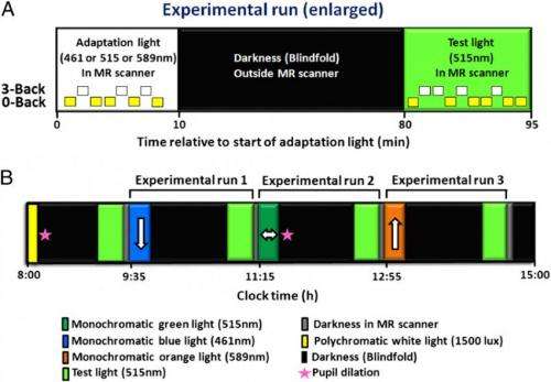
In the experiment, while continuously exposed to the same test light sixteen participants underwent identical, consecutive functional MRI recordings, during which they performed a simple auditory detection task and a more difficult auditory working memory task. "The idea here was to test the impact of the same test light on the cognitive brain responses to the exact same task," Vandewalle says. This allowed the scientists to isolate the determining variable – that is, the spectral nature of the previous light exposure to blue, green or orange that occurred one hour prior to the test itself. "We randomized the order of prior light to rule out an order effect, and also asked to participants how they felt in the MRI scanner." Importantly, he notes, no subjective difference on task difficulty or test light visual perception were found.
The researchers also hypothesized that based on the spectral sensitivity of melanopsin's two states, the impact of a given test light on cognitive brain responses would be increased, decreased, or intermediate after prior exposure to longer, shorter, or intermediate wavelength light, respectively. Cooper says that the long one-hour initial exposure was aimed at setting the sensitivity of melanopsin, relying on the assumption that during the 60-minute period of darkness until the cognitive test under green light, melanopsin would remain in a given state. "Previous studies in animals and on the biochemical properties of melanopsin have shown that while prior exposure to blue wavelength light reduces the sensitivity of photopigment to generate a response, and orange light increases sensitivity to generate a response, mid-wavelength green light is neutral between the two other wavelengths. We therefore chose green light as the test light condition."
Vandewalle and Cooper describe the two novel key strategies that the team developed to address these challenges.
- The first was to use of a continually modulated light intensity during the cognitive test phase, allowing them to simultaneously track brain activity modulation. Brain signals can constantly change, and this variation is a challenge for extracting the valid brain activity signals from random background noise. This approach allowed the scientists to retain brain signals that changed in a correlative way with the light signal, while rejecting other superfluous variations.
- The other key factor was to develop a mathematical model to predict the light spectrum to optimally drive melanopsin to the photoresponsive or the photoregenerative state. These two different states can rarely be measured directly, even in invertebrate bistable photopigments, and therefore must be derived.
Expanding on how their study demonstrates the importance of light for human cognitive brain function and support there being a cognitive role for melanopsin, Cooper notes that while it is generally accepted that light exerts stimulatory effects on the brain and increases performance on simple cognitive tasks, their paper presents two key findings: Firstly, that light specifically activates brain regions (such as the prefrontal cortex) involved in executive functions and improves cognitive task performance; and secondly, that the degree of activation depends on the spectral quality of a pre-exposure to light one hour earlier, with long wavelength orange light producing the most significant increase in brain activity in these cortical regions and short wavelength light inducing a relative decrease compared to orange light pre-exposure. "This is consistent with theories of a dual state photopigment, in which light serves as a primer to increase or decrease light sensitivity by converting the photopigment molecules to either the photosensitive or photoregenerative conformation," Cooper says. "Once this light-driven conversion occurs, the photopigment remains stable in a given conformation until a second light exposure to reactivate or deactivate the system. As our findings are consistent with this hypothesis, the results provide strong evidence that melanopsin is a major player in regulating the effect of light on cognitive responses. Moreover," he continues, "the current findings are also consistent with Vandewalle's previous study, demonstrating that in blind humans – that is, those lacking rods and cones but presumably with functional melanopsin – light stimulates brain activity during a cognitive task."
![Impact of the test light on executive brain responses depends on prior light. Orange blobs represent brain areas showing increased test-light impact after prior orange-light relative to prior blue-light exposure. Green blobs represent brain areas showing increased test-light impact after prior green-light relative to prior blue-light exposure (green arrows highlight these areas). Yellow blobs represent brain areas showing increased test-light impact after prior orange-light relative to prior green-light exposure (yellow arrows highlight these areas). Graphs show activity estimates of test-light impact on executive responses [3-back to 0-back; arbitrary units (a.u.); mean ± SEM] in the different brain areas after exposure to blue, green, and orange light. The numbers of the graphs correspond to brain locations on the central panels (as in Table 1). Graphs 1 and 2, left and right DLPFC; graph 3, left VLPFC; graph 4, left amygdala; graphs 5 and 6, left and right pulvinar; graph 7, substantia nigra; graphs 8 and 9, left and right fusiform gyrus; graph 10, cerebellum. *P < 0.05 corrected for multiple comparisons. Credit: Copyright © PNAS, doi:10.1073/pnas.1320005111 Thanks for the memories: Plant-like photopigment in the eye may play key role in human cognitive brain function](https://scx1.b-cdn.net/csz/news/800a/2014/cooperfig2.jpg)
The paper also contrasts the retinal pigment epithelium enzymatic retinoid cycle (required chromophore regeneration back to a light-sensitive state) with melanopsin being a dual-state photopigment in which photons drive both processes of phototransduction and part of chromophore regeneration. "As a dual state bistable photopigment," Cooper explains, "melanopsin chromophore regeneration does not require the retinoid cycle as do rods and cones. The phototransduction process is identical for rod-cone opsins and melanopsin: this begins with absorption of a photon that initiates the phototransduction cascade, causing the retinaldehyde chromophore to change from an 11-cis to an all-trans state, triggering a physiological response in the form of a change in membrane potential." 11-cis and all-trans states are isomers of retinaldehyde, a form of vitamin A.
In the all-trans state, says Cooper, the photoreceptor loses its capacity to respond to light – a state often referred to as photopigment bleaching – and requires conversion back to the 11-cis state. "This is where rod-cone opsins and melanopsin differ." Opsins are light-sensitive protein-coupled receptors. "Rods and cones achieve this through the light-independent enzymatic retinoid cycle, which is a slow process requiring roughly 15 minutes for cones and 30 minutes for rods. Humans that lack a functional pigment epithelium – for example, from mutations that cause retinitis pigmentosa – are incapable of regenerating the chromophore and are thus blind."
In melanopsin, however, chromophore regeneration is light dependent. Contrary to rods-cones, the all-trans state is light sensitive and absorption of a long-wavelength photon can actively drive conversion of the chromophore back to the 11-cis conformation, thereby regaining phototransduction properties in a rapid light dependent process. This light-driven switching between two states, known as bistability, is common for invertebrate and plant photopigments. "In a bistable photopigment such as melanopsin," Cooper points out, "both states are stable and can remain for some time in the same conformation in darkness, waiting for new photon availability. This is why we used a pre-exposure of light (blue, green or orange) to prime or drive the system to a given state and then allowed one hour of darkness. It's unknown how long melanopsin can remain in the same state, but certain bistable invertebrate photopigments remain stable for periods of hours to days." He notes that this does not exclude the possibility that melanopsin may also partly use the retinoid cycle pathways – a process that has only recently been shown for bistable fly rhodopsin.
The researchers also conclude that melanopsin may provide a unique form of photic memory for human cognition. "We refer to this unique form of photic memory due to that fact that melanopsin is stable over a certain length of time in either the 11-cis or all-trans state," Cooper tells Medical Xpress. "Once light sets the system to one of these states, melanopsin will remain in that state until another photon is absorbed. Accordingly, the spectral nature of previous light exposure determines the sensitivity to a subsequent light stimulation. In other words, melanopsin 'remembers' the most recent history of light exposure." Interestingly, Cooper notes that the term photic memory was first applied in the 1970s to plant photopigments – in particular pigments involved in stimulating or inhibiting seed germination – to express the idea, borrowed from electronics, that the photopigment acted as a bistable flip-flop, even though melanopsin time constants are unknown.
In fact, say Vandewalle and Cooper, melanopsin may play a broader role than previously thought, and has been implicated in regulating a surprisingly broad range of both acute and sustained non-visual responses to light – that is, non perceptive, unconscious light perception – including circadian and seasonal rhythms, hormonal secretion, gene expression, sleep/wake cycle, reproduction, cardiovascular function, cerebral blood flow, body temperature, acute heart rate, glucose metabolism, immune system function, and alertness. More recently, they note, melanopsin has been shown to participate in vision, perhaps by adjusting sensitivity to the overall light environment. "Our study now demonstrates that melanopsin participates in memory and cognitive functions, and considering these diverse functions, melanopsin may play the broadest functional role of any photopigment in the mammalian retina."
Cooper points out when melanopsin was first cloned in 1998 by Ignacio Provencio at the University of Virginia, Provencio showed that melanopsin has greater resemblance (in terms of amino acid sequence homology) to invertebrate photopigments than to vertebrate rod and cone opsins – a finding that has since been confirmed by others in phylogenetic analyses of animal photopigments. "This showed that melanopsin most closely resembles one of the fly rhodopsins – but strangely enough, melanopsin has not been shown to be present in any invertebrate." That said, he adds that there was debate over whether the rhabdomid or ciliary (that is, invertebrate or vertebrate) types of photopigments were more primitive and appeared first in evolution. However, it is now generally considered that both appeared at the same time and were present in the most primitive organisms – and modern detection techniques have been shown that many, even most, organisms have at least one form of both. Even then, the scientists add, while It has been known for some time that the previous light history effect – that is, light received several hours or days previous alters our sensitivity to continued light exposure – can in part be attributed to melanopsin, these effects also depend on a certain type of hysterisis of the circadian clock itself. (Hysterisis the dependence of a system on both its current and past environments,)
In terms of technological application, both Vandewalle and Cooper feel that their results are highly pertinent for the design of public and domestic lighting systems. "Ours and other findings strongly argue that light intensity, spectral quality and timing can be used to optimize non-visual or biological photic responses. A simple question, they say, is do we require that same amount and type of light at different times of the day? "This question can be addressed both from the points of view of the biological effects of light and the optimal use of energy consumption for lighting. Most chronobiologists would recommend high levels of blue light in the morning to help set our biological clock and stimulate waking and favor alertness. In contrast, light can also have deleterious effects if administered at inappropriate times. For example, in the evening it appears more logical to avoid the stimulatory effects of light and favor lower lighting conditions with long-wavelength dominated light that prepares for sleep onset. With the introduction of modern LED lighting technologies this has now become feasible."
They acknowledge that one difficult challenge is to reconcile the optimal lighting for biological effects or for visual perception. "It must be recalled that melanopsin is a basically blue light-sensitive photopigment, even through prior light exposures to orange light increase this response. In contrast, photopic sensitivity for visual perception is maximal in yellow light. Thus, future lighting schemes must take into account this dual function of the retina to increase or decrease visual and non-visual functions according to the desired outcome. Finally, our retinal light detection systems have evolved under, and are therefore adapted to, sunlight spectral and intensity changes occurring throughout the day. It thus seems obvious that use of natural light should also be an essential part of domestic and commercial lighting design."
Looking ahead, says Cooper, the research community needs next is to determine what specific cellular processes in the brain are up-regulated or down-regulated by light and darkness. "We now know what brain areas are involved, but our knowledge of the neurotransmitter systems and gene expression brought into play is still fragmentary. This would seem to be a fundamental question for biology and for the diurnal primates that we are. Ask any person on the street if he or she feels better on a sunny day or during the summer compared to winter and the response will be an overwhelming yes – yet biologists cannot yet provide all the answers. This is one aspect of our research," he concludes, "that we are currently pushing forward."
More information: Photic memory for executive brain responses, Proceedings of the National Academy of Sciences, Published online before print on March 10, 2014, doi:10.1073/pnas.1320005111
© 2014 Medical Xpress. All rights reserved.


