April 10, 2015 report
Sleep- and wake-dependent neuronal changes in fruit fly brains

(Medical Xpress)—The difficulty of studying sleep- and wake-dependent changes in neural activity in humans has led to comparative studies using animal models. Though MRI technology allows researchers to study macro-level brain responses across groupings of thousands of neurons in humans, engaging with changes at the level of individual neurons is trickier, as the brain is encased in a skull.
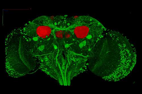
Drosophila melanogaster—the common fruit fly—provides a good model for comparative study, because its neural characteristics are broadly analogous to those of rodents and humans. Additionally, its brain cells can be more easily monitored via two-photon microscope.
A group of researchers from the department of psychiatry at the University of Wisconsin-Madison conducted a study to determine if the effects of sleep and wake had any effect on calcium levels in the Kenyon cells of fruit fly brains. Though previous research recorded GCaMP fluorescence in fruit fly brains, the effects of sleep and wake cycles had not been studied. They published their results in the Proceedings of the National Academy of Sciences.
But can fruit flies sleep while tethered to a microscope?
It was first necessary to determine whether immobilized flies could fall asleep under a two-photon microscope. They demonstrated that when the flies became visibly inactive for a period greater than five minutes, their ability to behaviorally respond to a stimulus was reduced. To stimulate putatively sleeping flies, the researchers switched on the microscope's laser light. Flies that had been in quiescent periods of greater than five minutes were far less likely to respond to the stimulus than flies whose quiescent periods were interrupted with stimulation before five minutes had elapsed.
They also found that sleep as defined by this study was homeostatically regulated by the duration of the previous wake cycle, consistent with previous results. "Thus, we conclude that the state of quiescence as defined in our experimental setup qualifies as sleep and not simply rest," the researchers write.
Changes during sleep
The researchers were interested in studying the changes in neuron calcium levels, as measured by GCaMP5 fluorescence, while the fly was transitioning between periods of wake to sleep and back. They found that calcium levels in individual neurons increased when flies woke up and decreased when they fell asleep.
In addition to the study of spontaneous activity during sleep and wake cycles, they measured evoked activity in the animals' appendages by exposing the flies to stimuli—specifically, exposure to oxygen and vinegar. As expected, these evoked activities were diminished in sleeping flies and higher in those that were awake. "A decline in the electrical response of neurons to different kinds of stimuli is a classical marker of sleep, documented in mammals as well as bees," the researchers write. "Thus, we were able with calcium imaging to detect sleep/wake-dependent changes in both spontaneous and evoked activity of Kenyon cells."
But what about history-dependent effects?
Looking through the data, the researchers found that flies that had previously been awake for five to eight hours prior to testing showed more bright cell bodies before the vinegar odor stimulus than flies that slept before testing. This result is consistent with with previous findings in mammal studies that spontaneous neural activity increases with time spent awake, decreasing after sleep.
After long periods of wake, however, the observed Kenyon cells were unable to respond consistently to repeated exposure. Comparing the results between two sets of trials, they determined that it was unlikely to be due to habituation to the stimulus; rather, the results corresponded with previous study results demonstrating that sleep may not be a global phenomenon in the brain—in sleep-deprived rats, for instance, it is known that small groups of cortical neurons go offline while the rest of the brain is in a wake state. This phenomenon likely explains cognitive deficits that occur in sleep-deprived humans.
More information: "Sleep- and wake-dependent changes in neuronal activity and reactivity demonstrated in fly neurons using in vivo calcium imaging." </>PNAS 2015 ; published ahead of print March 30, 2015, DOI: 10.1073/pnas.1419603112
Abstract
Sleep in Drosophila shares many features with mammalian sleep, but it remains unknown whether spontaneous and evoked activity of individual neurons change with the sleep/wake cycle in flies as they do in mammals. Here we used calcium imaging to assess how the Kenyon cells in the fly mushroom bodies change their activity and reactivity to stimuli during sleep, wake, and after short or long sleep deprivation. As before, sleep was defined as a period of immobility of >5 min associated with a reduced behavioral response to a stimulus. We found that calcium levels in Kenyon cells decline when flies fall asleep and increase when they wake up. Moreover, calcium transients in response to two different stimuli are larger in awake flies than in sleeping flies. The activity of Kenyon cells is also affected by sleep/wake history: in awake flies, more cells are spontaneously active and responding to stimuli if the last several hours (5–8 h) before imaging were spent awake rather than asleep. By contrast, long wake (≥29 h) reduces both baseline and evoked neural activity and decreases the ability of neurons to respond consistently to the same repeated stimulus. The latter finding may underlie some of the negative effects of sleep deprivation on cognitive performance and is consistent with the occurrence of local sleep during wake as described in behaving rats. Thus, calcium imaging uncovers new similarities between fly and mammalian sleep: fly neurons are more active and reactive in wake than in sleep, and their activity tracks sleep/wake history.
© 2015 Medical Xpress


